- Common injury
- Anatomically thin part of lower humerus
- Mechanism: Fall on outstretched hand - Hyper-extension of elbow (most common type) (impact against olecranon)
Complications and Assessment
Needs immediate care
-
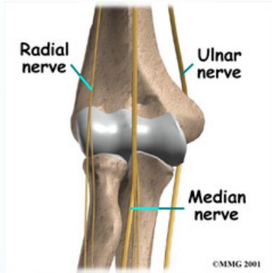
Nerve injury: Median, Radial
- Test: Oppose thumb with little finger
- Test: Flex/extend fingers/wrist
-
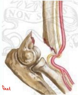
Vascular injury: Brachial artery
- (tenting, kinking) - only by touching the artery the patient feels spasm in distal part of forearm
- Assessment:
- Palpate brachial & radial pulses
- Check capillary refill in fingers (should be normal)
-
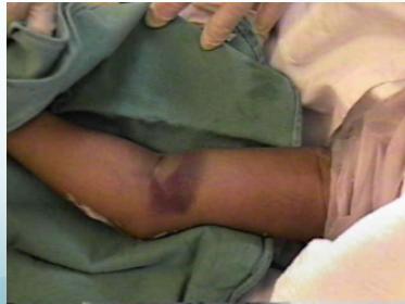
Swelling:
- Can lead to compartment syndrome → Volkmann’s ischemia
- A real emergency - Permanent damage possible
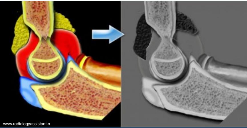
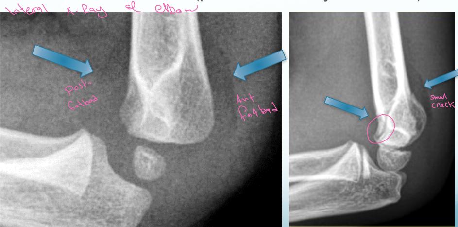
Fat Pad Sign:
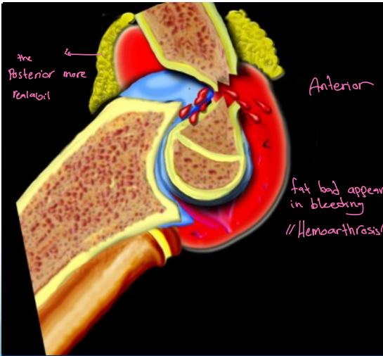
- Posterior fat pad always appears in bleeding/hemoarthrosis - always abnormal
- Anterior fat pad may appear in bleeding
Important: If there’s no fracture line but there’s fat pad, treat it as minimally displaced with back slab for 2-3 days and repeat X-ray
X-Ray Lines in Elbow
Lateral elbow X-ray image
- Fat pad sign:
- Indicates a fracture (posterior is always abnormal)
Anterior Humeral Line:
- A line drawn on a lateral view along the anterior surface of the humerus should pass through the middle third of the capitellum
Undisplaced :
- Treat with above elbow cast, elbow 90o for 3 wks.
- Follow-up x-rays after 1 week to document alignment
- X-rays at 3 weeks – if callus noted, discontinue cast & start active ROM
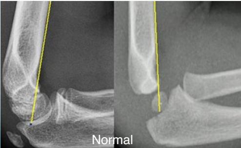 Normal
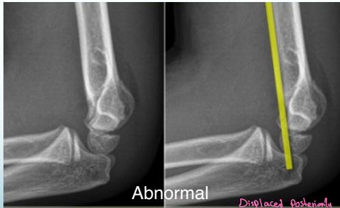 Abnormal - Displaced posteriorly
Treatment
- Immobilize elbow before radiographs to avoid further injury from sharp fragments
- Flexion 30° = least nerve tension
Undisplaced fractures:
- Long arm Bivalve cast only
- Forearm neutral, elbow 90° for 3 weeks
- Follow-up x-rays after 1 week to document alignment
- X-rays at 3 weeks – if callus noted, discontinue cast & start active ROM
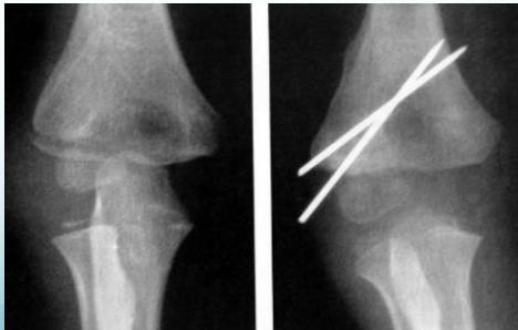
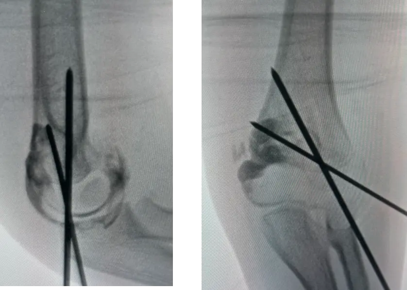
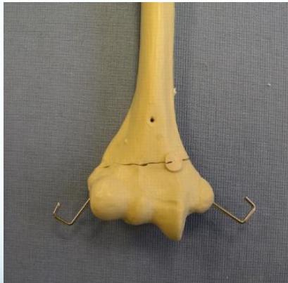
Displaced fractures:
- Closed reduction and fixation with K-wires & slab
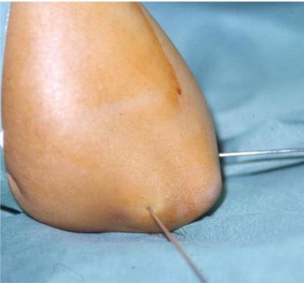
Case Example: 6-year-old girl, fell from swing
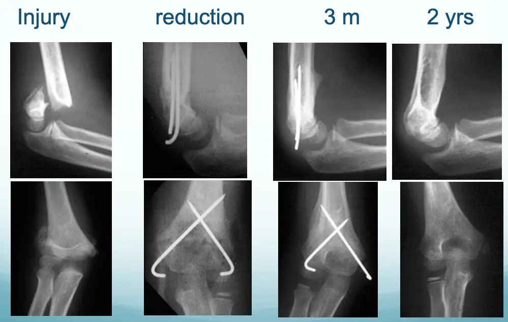
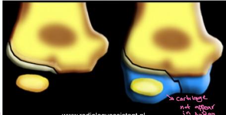
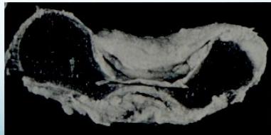
Lateral Condyle - Humerus
- Mostly cartilaginous
- Intra-articular fracture
- Fracture easily missed
- Displacement not appreciated/underestimated
- Needs fixation even if undisplaced
- Treatment: reduction, K-wire, slab
- If not fixed, may displace