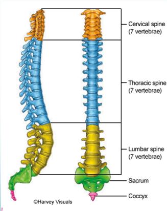
Vertebral Segments
- 7 Cervical vertebrae
- 12 Thoracic vertebrae
- 5 Lumbar vertebrae
- 5 Sacral vertebrae (fused)
- 4 Coccygeal vertebrae (fused)
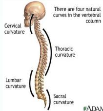
Natural Spinal Curves
The spine has 4 natural curves to distribute mechanical stress during movement:
- Cervical: Lordosis (inward curve)
- Thoracic: Kyphosis (outward curve)
- Lumbar: Lordosis (inward curve)
- Sacral: Kyphosis (outward curve)
Note: When viewed from the front or back, the spine should appear straight.
Intervertebral Disc Structure
The disc is composed of:
- Nucleus pulposus - Soft, gel-like center
- Anulus fibrosus - Tough, rigid outer ring (حلقة ليفية)
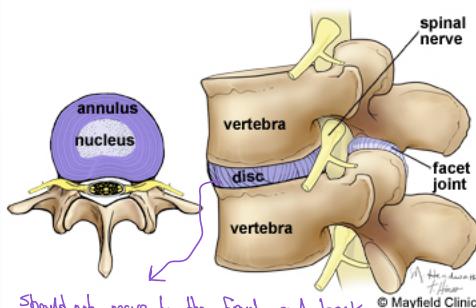
Important: The disc should not move anteriorly or posteriorly as this may injure the spinal cord, causing:
- Compression of nerve root or spinal cord
- Sciatica symptoms
Common Symptoms
- Pain - Primary presenting symptom
- Stiffness / loss of function - Common in elderly patients with osteoporosis
- Deformity - More common in children
- Neurological symptoms:
- Numbness, paresthesia (abnormal sensation)
- Hyperesthesia (increased sensitivity)
- Hypoesthesia (decreased sensitivity)
- Muscle weakness
- Sphincter control problems (Bladder, Anus) - Cauda equina syndrome
- Quadruplegia (in severe cases)
Radiating Pain Patterns
- Brachialgia: Pain radiating through the Brachial Plexus from the neck to the upper limbs
- Sciatica: Back pain radiating to the lower limbs
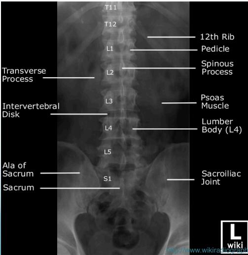
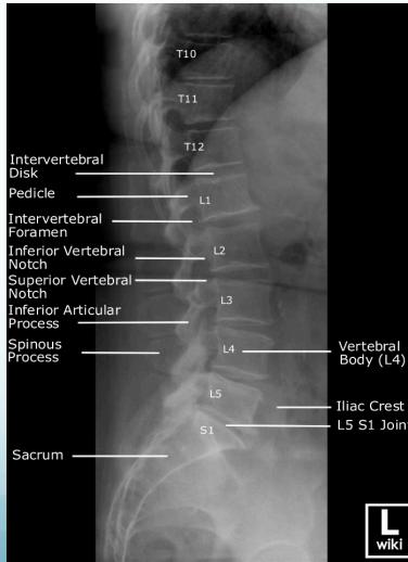
Imaging for Spinal Disorders
Radiographic (X-Ray) Views
- Standard Views: AP (Anteroposterior), LAT (Lateral)
- Specialized Views:
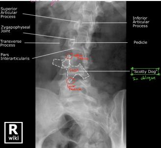
- Oblique: 45° angle to see structures between foramen
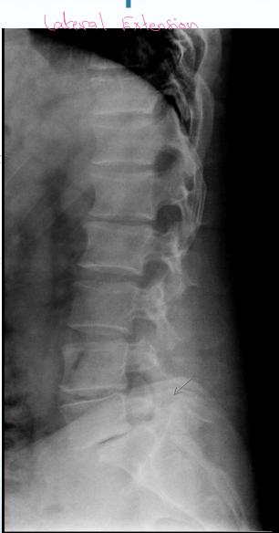
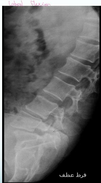
- LAT Flexion-Extension: To diagnose instability (especially in lower back pain)
- Open mouth: To visualize odontoid process
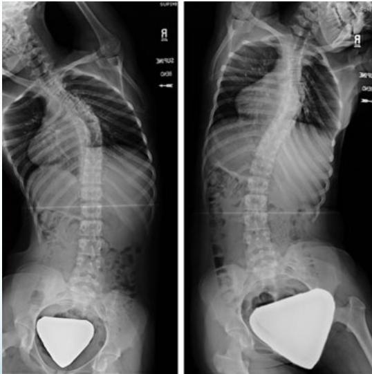
- Lat bending: For deformity assessment
- Deformity series: (e.g., scoliosis series)
Anatomical Structures Visible on Oblique View
- Superior Articular Process
- Zygapophyseal Joint
- Transverse Process
- Pars Interarticularis
- Inferior Articular Process
- Pedicle
“Scotty Dog” sign is visible on oblique radiographs
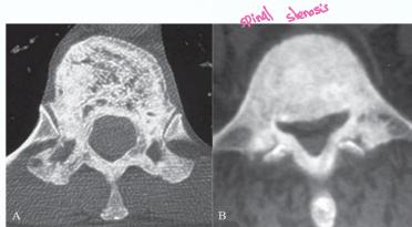
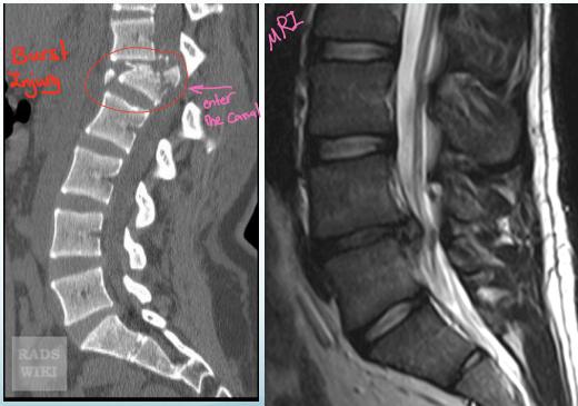
Advanced Imaging Modalities
- CT Scan: Shows bony structures in detail
- MRI: Shows soft tissues, discs, and spinal cord (gold standard for disc pathology)
- Other studies: Ordered as clinically indicated (e.g., PFT)