Most renal masses are detected incidentally on imaging performed for another indication or to evaluate nonspecific symptoms.
Preferred imaging exam: CT or MRI abdomen (with IV contrast)
CT abdomen (w/ IV contrast; multiphase renal protocol):
-
(Lesion(s) are often heterogeneous, with a combination of solid, cystic, necrotic, and/or hemorrhagic areas with thickened irregular walls, calcification, and variable enhancement.
-
Distorted renal outline
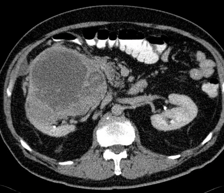
Axial CT of the abdomen after oral and IV contrast medium administration
A large, relatively sharply demarcated tumor can be seen in the right kidney. The CT shows a central, hypodense, necrotic area, which is surrounded by a solid, contrast enhanced, capsular region . This finding is characteristic of renal cell carcinoma.
