Table of Contents
- Development of the Reproductive System
- Key Components
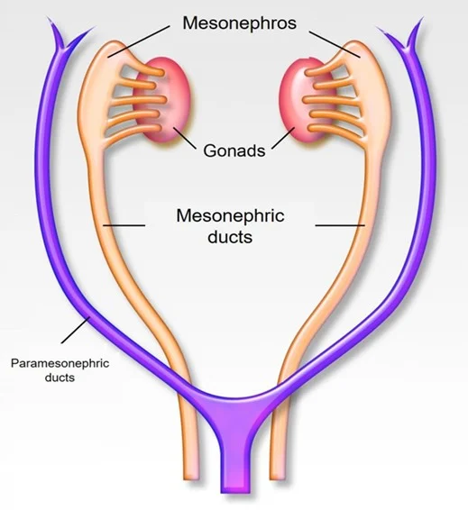
- Indifferent Gonads and Duct Systems (referencing
embryology-ps4-2.webp)- Differentiation Pathways
- Primordial Germ Cells (PGC)
- Male Gonads
- Female Gonads
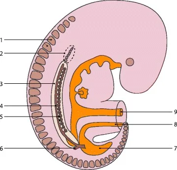
- Related Image Labels (referencing
embryology-ps9-1.webp)
- Related Image Labels (referencing
- Overview of Sexual Differentiation
- Sexual Differentiation Pathways
- XY Pathway
- XX Pathway
- External Genitalia and Urogenital Differentiation
- Male:
- Female:
- Uterine Anomalies
- Thank You!
Embryology
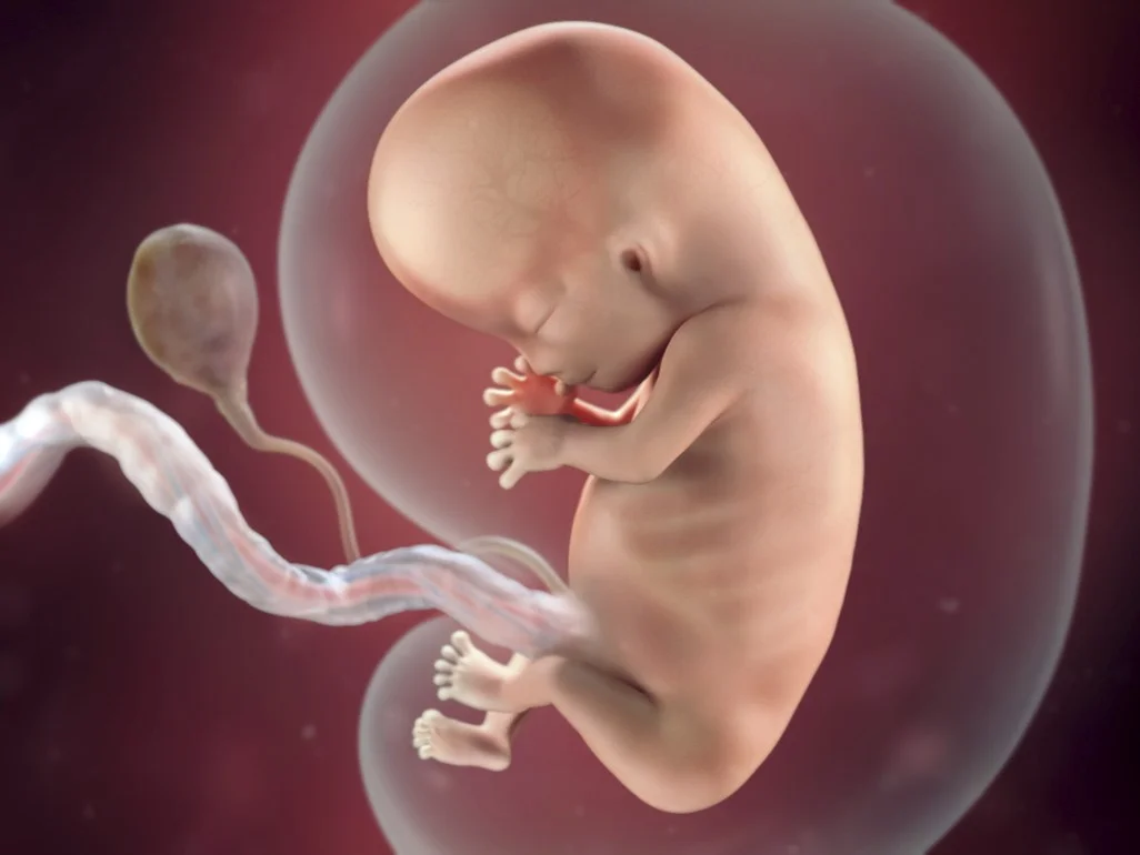
Development of the Reproductive System
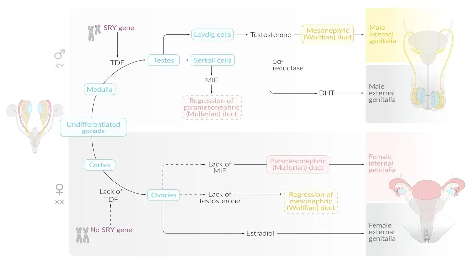
Development of the reproductive system begins with:
- Formation of undifferentiated gonads.
- and mesonephric and paramesonephric ducts.
- Further differentiation of the gonads:
- In male
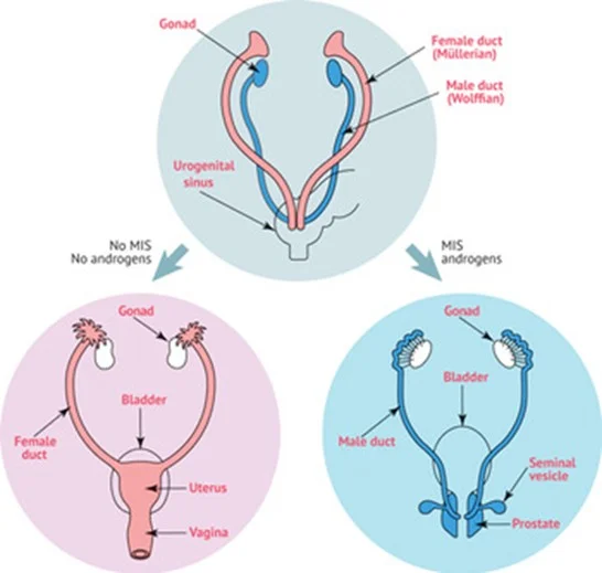
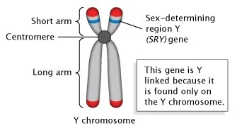
- SRY gene on the Y chromosome ➡️ testes.
- Testosterone (by Leydig cells) ➡️ differentiation of the mesonephric ducts into male internal sex organs.
- Dihydrotestosterone ➡️ differentiation of male external genitalia.
- MIF (by Sertoli cells) ➡️ suppresses paramesonephric differentiation.
- In male
The male and female reproductive tracts are derived from the same embryonic/fetal tissue.y
The gonads develop from three sources:
- Mesothelium (coelomic epithelium) lining the posterior abdominal wall
- Mesenchyme (intermediate mesoderm)
- Primordial germ cells
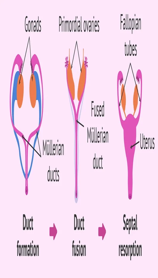
Up to the 7th week the internal genital organs in both sexes on both sides consist of two canal systems.
- The indifferent gonads consist of medulla and cortex.
- In XX: Ovary will originate from the cortex, and medulla will regress.
- In XY: Testes will develop from medulla and the cortex regress.
Cortex is the outermost.
Medulla is the innermost.
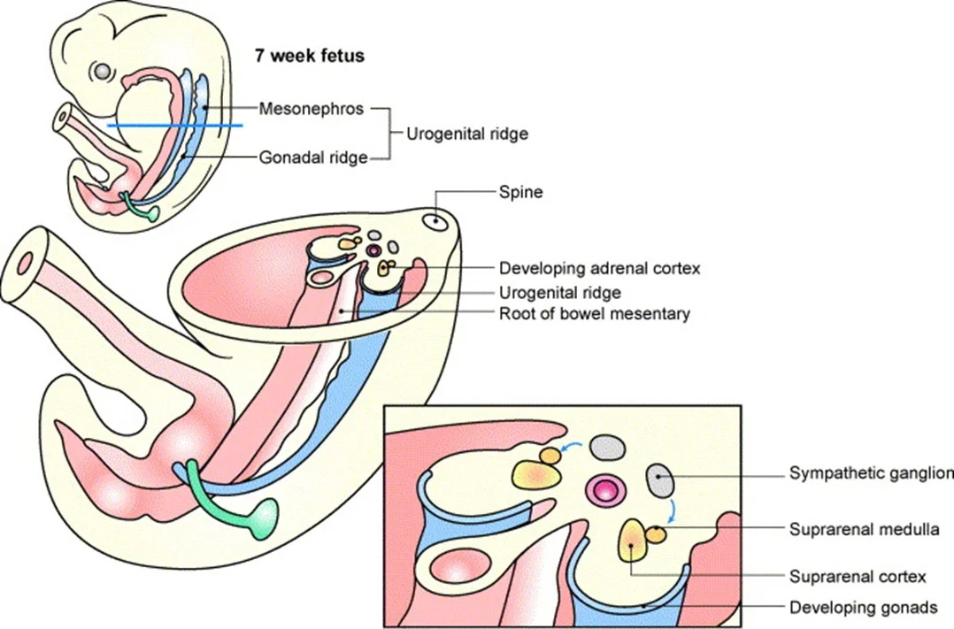
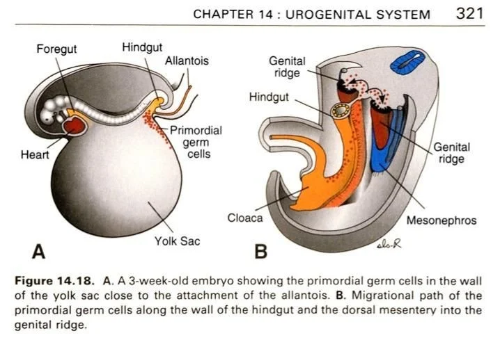
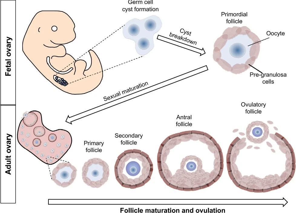
Primordial Germ Cells (PGC)
- Migrate from the allantois to the endoderm of the yolk sac.
- Then reach the genital ridge where they settle and lose their mobility.
Male Gonads
Female Gonads
- By 12th week
- The ovary develops more slowly than the testes.
- The somatic cell will develop into follicle cells in the absence of SRY gene. As there are no Sertoli cells, there is no AMH.
- Stromal cells will surround the PGCs.
- The germ cells go on to differentiate into oogonia, proliferate, and enter the first meiotic division to form primary oocytes.
The maximum number of primordial follicles is reached at 20 weeks’ gestation when there are six to seven million primordial follicles present.
- The numbers of these reduce by atresia; they are 1-2 million at birth and 400,000 at puberty.
The fusion of the Mullerian ducts forms:
- the uterovaginal canal that gives rise to the
- ✓ Uterus
- ✓ Cervix.
- ✓ upper two thirds of the vagina.
- The lower third of the vagina urogenital sinus.
- The embryological origin of the hymen is controversial.
- The unfused, cranial portions of the paramesonephric ducts become the Fallopian tubes.
Many anomalies related to development of the uterus and vagina are attributable to abnormal fusion or regression of the caudal portion of the paramesonephric duct.
Overview of Sexual Differentiation
- Weeks 1–6: No sexual differentiation is apparent (no phenotypical differences).
- Week 6–7: Differentiation of the mesonephric and paramesonephric ducts begins.
- Week 12: Development of the external genitalia is complete.
- Week 20: Phenotypic differentiation is complete.
- Week 33: Descent of the gonads is complete.
Sexual Differentiation Pathways
Male: testosterone → dihydrotestosterone (via 5α-reductase) → differentiation of embryonic structures into male external genitalia.
Female:
estradiol and absence of DHT → differentiation of embryonic structures into female external genitalia
Uterine Anomalies
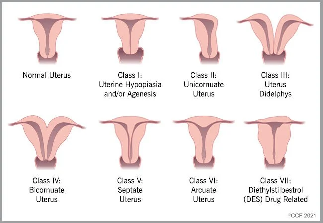
- Normal Uterus
- Class I: Uterine Hypoplasia and/or Agenesis
- Class II: Unicornuate Uterus
- Class III: Uterus Didelphys
- Class IV: Bicornuate Uterus
- Class V: Septate Uterus
- Class VI: Arcuate Uterus
- Class VII: Diethylstilbestrol (DES) Drug Related
°CCF 2021
Thank You!