Case 1 Z
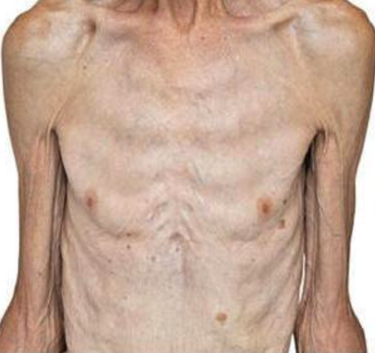 Patient with weight loss and diarrhea. Suspected of having malabsorption syndrome.
++ Female patient with diarrhea
Biopsy show: destructive intestinal mucosa..
And 20g. lipid in stool
-
Name any 2 etiologies of fat malabsorption? Celiac disease, pancreatitis.
-
Which serologic test will you do for the diagnosis of celiac disease? Serum anti-gliadin and anti-endomysial antibodies.
-
Name any imaging study which is used in the workup of malabsorption? CT Abdomen, Endoscope with biopsy.
Case 2
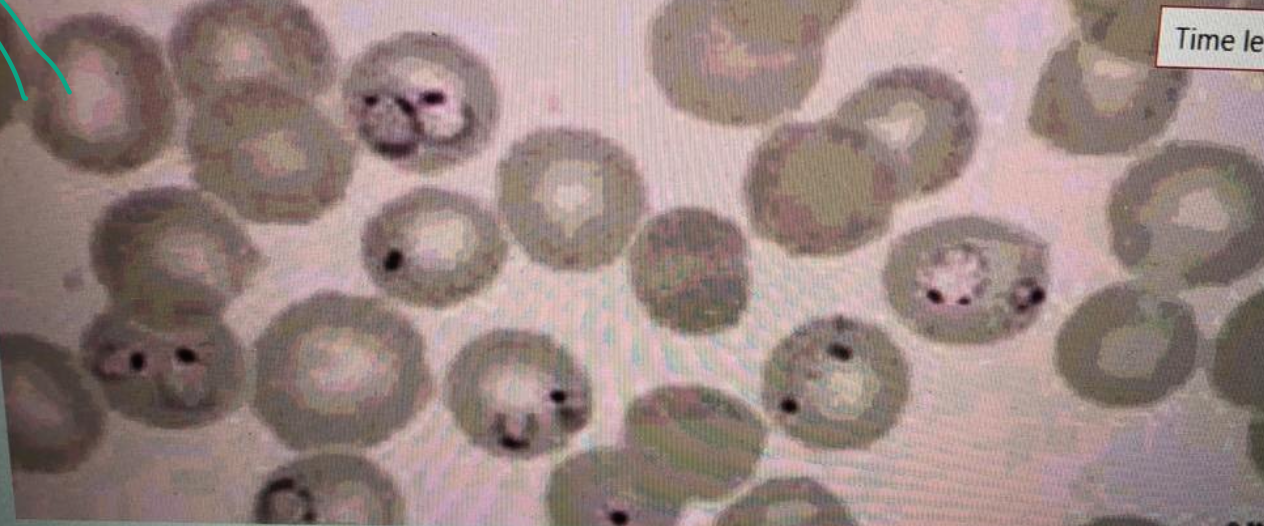 A 20-year-old male comes with fever since 5 days. Fever is episodic. The peripheral blood film is shown below.
- What is your diagnosis based on the abnormality on the slide?
- Malaria.
- Outline 2 causes of anemia in this infection?
- Mechanical destruction of RBCs, reduced erythropoiesis in the bone marrow.
- Outline any 3 features of severe form of this infection? Z
- Cerebral malaria, blackwater fever, hypoglycemia
- Name any 2 medicines used to treat this infection, and one of them used during pregnancy?
- Hydroxychloroquine (Pregnancy), mefloquine.
Case 3
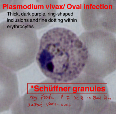 Thick, dark purple, ring-shaped inclusions and fine dotting within erythrocytes.
“Schüffner granules” (very specific if seen in blood film, suspect vivax-oval)
- Name the cause: Vivax - Oval
- Treatment: Primaquine
- What is the specific name? Schüffner granules
Case 4
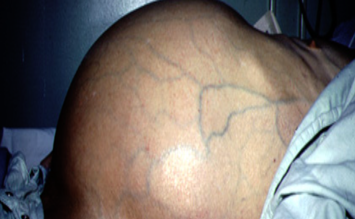 Ascites in liver cirrhosis
findings:
- Skin pigmentation
- Abdominal distention
- Visible vessels
Lab test:
- Saag
- Chemistry
- LFT
- U/S
Q&A
- Causes: Liver Cirrhosis - CHF
- Infectious Complication: Spontaneous Bacterial Peritonitis
- What is SAAG? Serum Albumin to Ascites Albumin Ratio
- Diagnosis: Ascites
Case 5
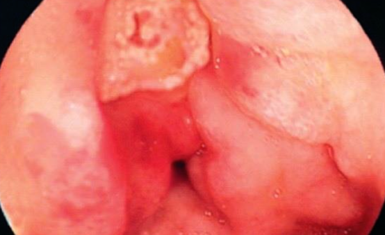 25 year old patient. Complaining of epigastric pain and dyspepsia since 1 month. Upper endoscopy showed duodenal ulcer as shown & H.pylori test was positive.
-
Name any 2 investigations used to diagnose H. pylori? Stool antigen test Urea breath test Z
-
Which combination of medicines is used to treat H. pylori infection?
- PPI’s - Omeprazole
- Clarithromycin
- Amoxicillin
-
Name any 1 complication of duodenal ulcer? Perforation
Case 6
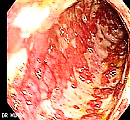 This is an endoscopic image of colon affected by ulcerative colitis. The mucosa is blotchy and broken in many places.
Q1: Name 3 common clinical symptoms of ulcerative colitis?
Lower abdominal pain, diarrhea, blood and mucus with the stool.
Q2: Name 2 features of a severe episode of UC?
Leukocytosis, increase frequency of the stool more than 6 times in the day, anemia.
#Z
Q3: Name 2 ttt options for UC?
5-ASA, Corticosteroids.
#Z
Q4: Name 3 extracolonic manifestations of UC?
Arthritis, Ancylosing spondylitis, Uveitis, Episcleritis
CASE 7
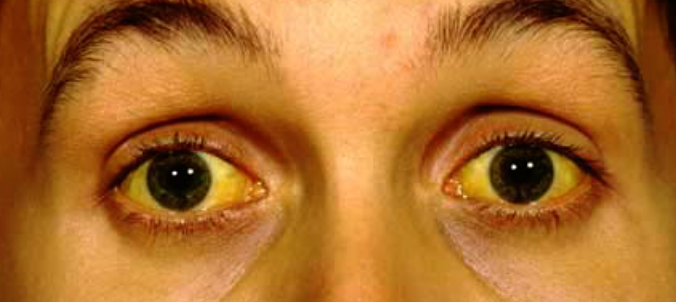 Q1: JAUNDICE
Q2: Mention 4 risk factors. Hemolytic Anemia. Alcohol - drugs: Paracetamol - GB stones
Q3: 4 Signs.
- Yellowish skin
Q4: Investigations. Serum Bilirubin, Urine Bilirubin, LFT
Q5: Treatment. Treat the cause
Case 8

A 47 year old woman complains of generalized tiredness, low mood, altered bowel habits and more recently generalized itching. O/E she has 4 cm hepatomegaly and xanthelasma around her eyes.
Her liver function tests show:
- Total bilirubin 60 μmol/l
- AST 35 IU/l
- ALT 34 IU/l
- ALP 180 U/l
- GGT 90 U/l
- Albumin 39 g/l
- AMA titer 1:140
-
Describe the abnormalities present?
a. Increases bilirubin, ALP, GGT, AMA -
Suggest 2 possible causes of this diagnosis? Z
1- Primary Biliary Cirrhosis
2- sclerosing cholangitis
Case 9
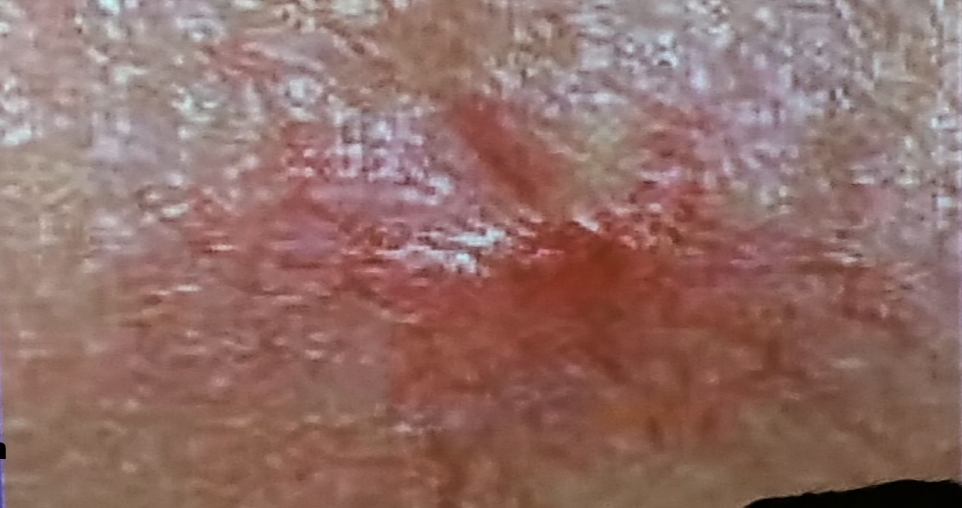 This lesion was found on routine medical examination of military recruit who was fit and has been medically free.
-
What is the lesion?
a. Spider-angioma -
Name 2 clinical conditions in which this lesion may be seen?
1-Cirrhosis (partial-HTN)
2-increase portal hypertension #Z -
What advice will you to this person?
a. Reassure
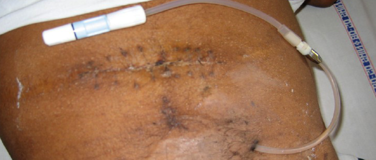
Peritoneal dialysis catheter
A 43 year old woman presented with diarrhea
The following results were obtained:
3 day fecal fat 30 mmol/day
Jejunal biopsy subtotal villous atrophy
#Z
-
What is the most likely diagnosis?
Celiac Disease -
What treatment is recommended?
Gluten-free diet