Q&A RESP & CVS IMG
Basic investigations for chest
1) Plain film=chest x-ray(CXR)
(you can detect Pneumonia , TB and Bronchiectasis)
2) CT (computed Tomography):
used it if you want to visualize the mediastinal structures, if you suspect a mass or details of lung parenchyma
- HRCT (high resolution CT).
- CT with contrast.
- CT angiography
3) MRI ( limited use)
4) Ultrasound
5) Radioisotopes studies
6) Angiography
Signs of Respiratory & Cardiovascular findings
abdominal conditions present as chest disorder
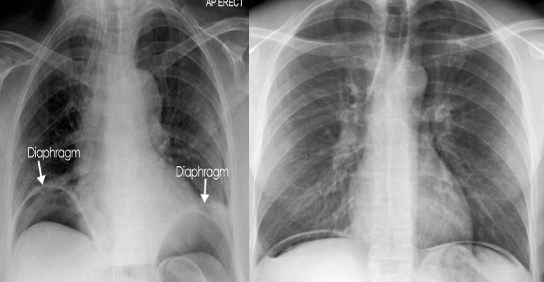
- Air under the domes of diaphragm in pneumoperitoneum.
- It occur in perforation of bowel

Respiratory Findings
- Pneumonia
- Pleural Effusions
- Pneumothorax
- Lung Collapse
- PNEUMONECTOMY
- Emphysema
- Bronchiectasis
- Lung Abscess
- Spherical opacities (lung mass & nodule)
- Thymoma
- Pulmonary Tuberculosis
- Hilar Nodes Enlargement
- Respiratory distress
Cardiovascular Findings
- Dextrocardia
- Pericardial effusion
- Mitral Stenosis
- Coarctation of the aorta
- Aortic Dissection
- Pulmonary Embolism
- Pulmonary Edema
Further ridings Reference Book (Andrea G. Rockall, 7th edition) - •Page 40 – 44 Spherical opacities (lung mass, lung nodule) To Multiple pulmonary nodules