Ultrasound can detect intra-abdominal fluid and assess the bowel wall in certain situations, but gives limited information about the bowel mucosa
Ultrasound is used for the diagnosis of infantile pyloric stenosis, Intussusception and in cases of suspected Appendicitis when the diagnosis is not obvious clinically
Hepatic Ultrasound:
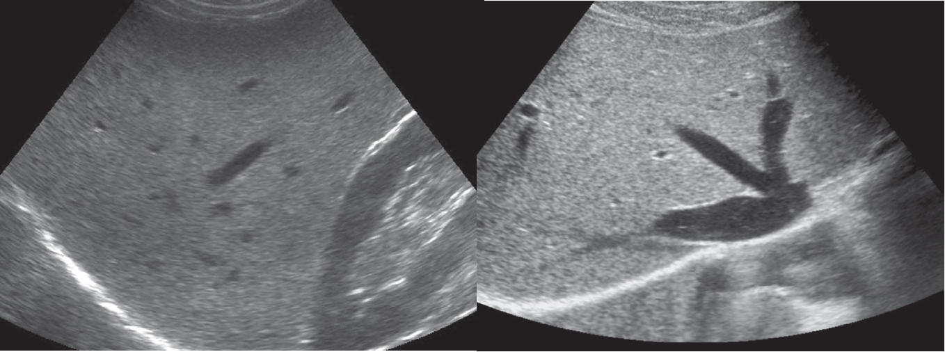
The normal hepatic parenchyma is of uniform echogenicity, composed of medium and low level echoes, interspersed with the bright echoes of the portal triads and echo-free areas corresponding to large hepatic veins.
 1st Ultrasound of normal liver showing a uniform echo pattern interspersed with bright echoes of portal triads and echo-free areas of hepatic and portal veins
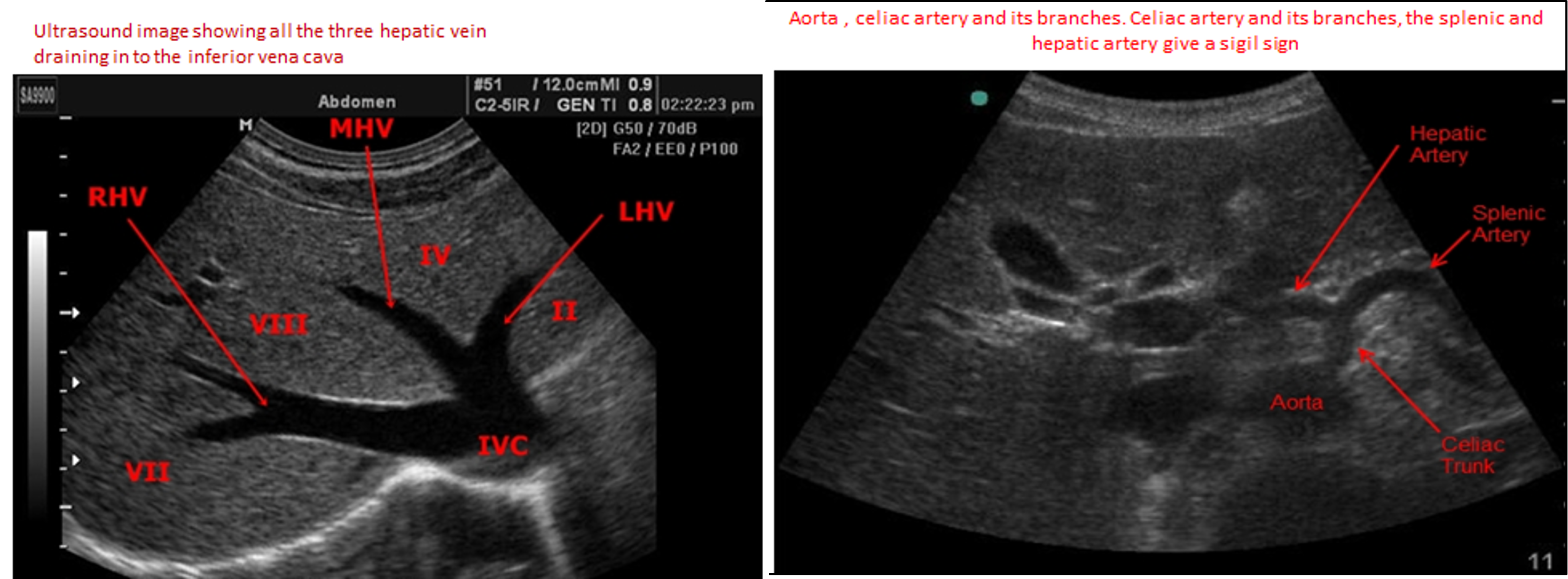
2nd Ultrasound of normal liver. showing the right , middle and left hepatic veins draining into the inferior vena cava as it penetrates the diaphragm.
BILIARY SYSTEM
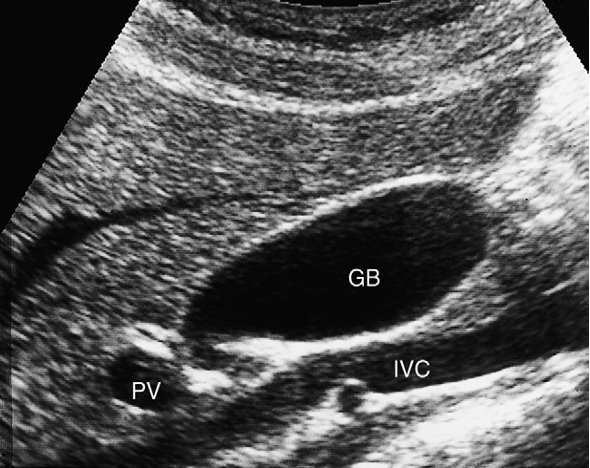
Ultrasound is the initial investigation for demonstrating the GB and bile ducts. , it is the next best step for patients with suggestive clinical features, risk factors for cholelithiasis, and cholestatic.
The common bile duct is seen as a small, tubular structure lying anterior to the portal vein in the porta hepatis and should not measure more than 7 mm in diameter unless the patient has had a cholecystectomy, when it may be larger.

 Transverse Plane
The transverse sectional images are presented in descending order from the dome of the diaphragm to the umbilicus.
Transverse image at the dome of the liver shows left and middle hepatic vein draining into the inferior vena cava. The homogeneous liver texture is well seen
 - Sigil/// = Seagull sign
