Indication:
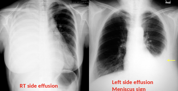
- Standard initial imaging modality for detecting pleural effusion.
- Lateral decubitus view (most sensitive): allows for detection of fluid collections as small as 5 mL
Supportive Findings:
- Typically unilateral blunting of the costophrenic angle
- Homogeneous density with a meniscus-shaped margin (meniscus sign)
- Large effusion — Complete opacification of the lung — Mediastinal shift — Tracheal_deviation away from the effusion (space-occupying lesion)


Computed tomography z
-
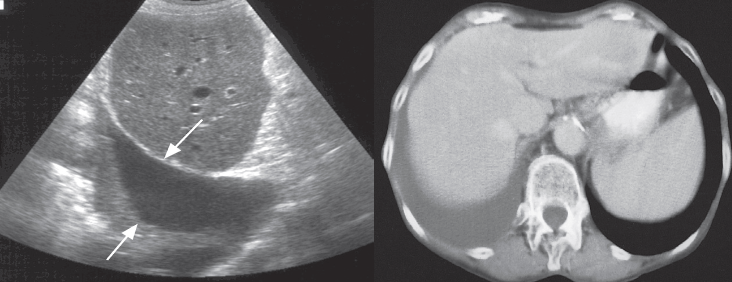
Pleural effusions are usually seen as an area of homogeneous fluid density between the chest wall and lung. CT can be used to distinguish between lung abscess and empyema. CT can be used to direct the placement of drainage tubes.
Ultrasound Z
-
Ultrasound is a simple method of determining whether pleural fluid is present and is a convenient method of imaging control to guide procedures such as pleural fluid aspiration or drainage. Pleural fluid can be recognized as an anechoic area between the chest wall and the lung
The split-pleura sign z
-
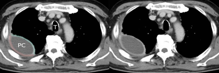
refers to thickening and contrast enhancement of the parietal and visceral pleurae separated by empyema or exudative effusion. The sign is considered reliable in distinguishing empyema from lung abscess.
-
Lung abscesses, furthermore, tend to be **round in contrast to the lenticular shape typical of empyemas.
CT chest (with contrast; axial plane; mediastinal window)
-
CT scan shows right-sided pleural fluid collection (PC). The collection has a lenticular (biconvex) shape and is accompanied by thickening of the adjacent parietal (red line) and visceral pleurae (green line).
