X-rays (Radiograph)
Often the first diagnostic imaging test, quick and cheap. Can detect:
- Spinal alignment and curvature
- Spinal instability – with flexion and extension views
- Congenital (birth) defects of spinal column
- Fractures caused by trauma
- Moderate osteoporosis
- Infections
- Tumors
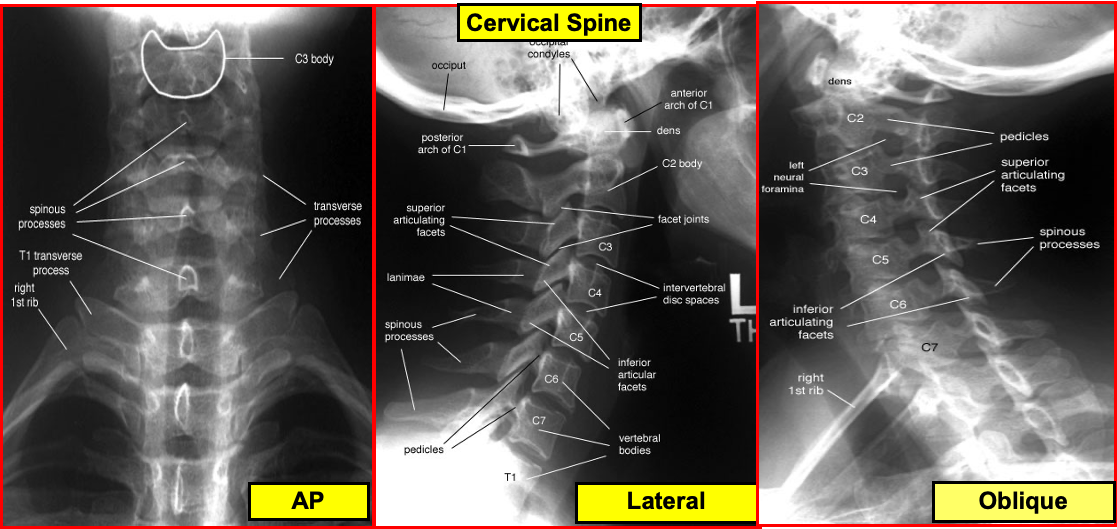
Cervical Spine
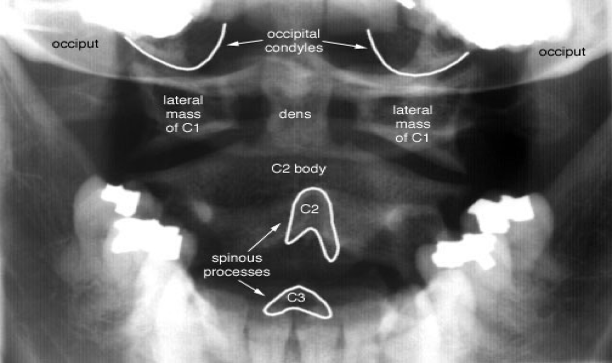
Radiographic evaluation Open mouth odontoid
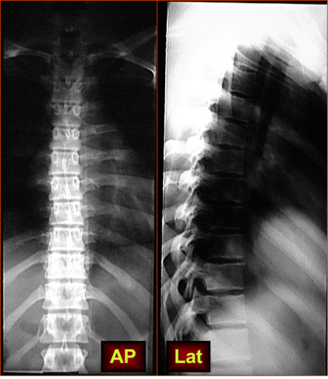
Thoracic Vertebrae
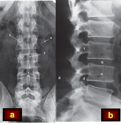 (a) Frontal view.
1, pedicles; 2, spinous process; 3, facet joint; 4, transverse process.
(b) Lateral view. 1, pedicles; 2, spinous process; 3, facet joint; 4, vertebral body; 5, disc space.
Note how the height of the disc spaces increases from L1 to L5 with the exception of the L5/S1 disc space which is normally narrower than the one above.
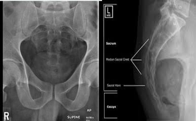
The Sacrum and coccyx
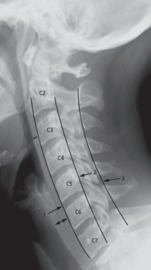
Normal cervical spine (CC TB)
showing lines to check alignment.
Line 1: runs along the anterior border of the vertebral bodies and corresponds to the anterior longitudinal ligament.
Line 2: runs along the posterior border of the vertebral bodies (posterior longitudinal ligament).
Line 3: runs along the junction of the laminae and spinous processes. (Spinolaminar line)
Line 2 & 3: Line 2 indicates the anterior extent and line 3 the posterior extent of the spinal canal.
There is a normal soft tissue distance between the anterior border of the spine and the posterior border of the airway (double-headed arrows)
