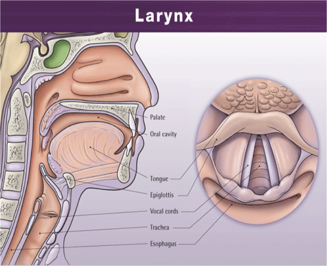
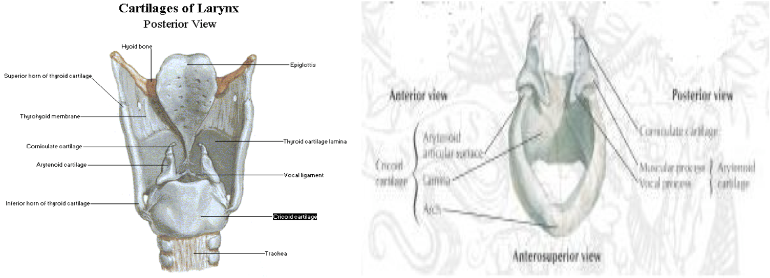
Skeleto-membranous Framework of Larynx
Components
- Thyroid cartilage
- Cricoid cartilage
- Paired arytenoids cartilage
- Epiglottis
- Hyoid bone
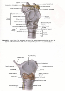
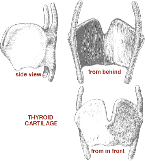
Thyroid Cartilage
- Shield-like structure
Cricoid Cartilage
- Signet ring shaped.
- The only complete skeletal ring for the airway.
- Both thyroid and cricoid cartilage are hyaline and prone to calcification.
Cricothyroid Joint
- Synovial joint with hinge motion.
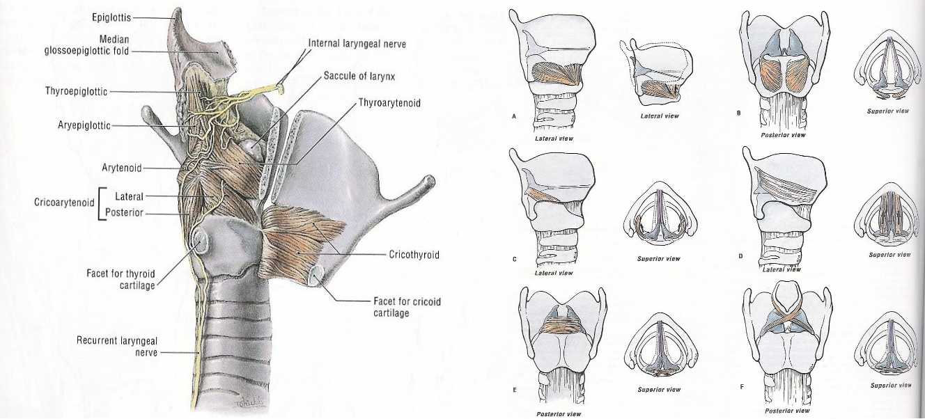
Arytenoid Cartilage
- Pyramidal shaped
- Apex, vocal & muscular process.
Cricoarytenoid Joint
- Synovial joint with rocking motion.
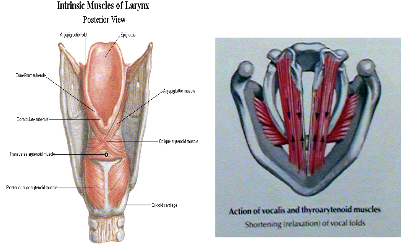
Corniculate and Cuneiform Cartilage
Epiglottic Cartilage
- Leaf-like structure
- Elastic cartilage
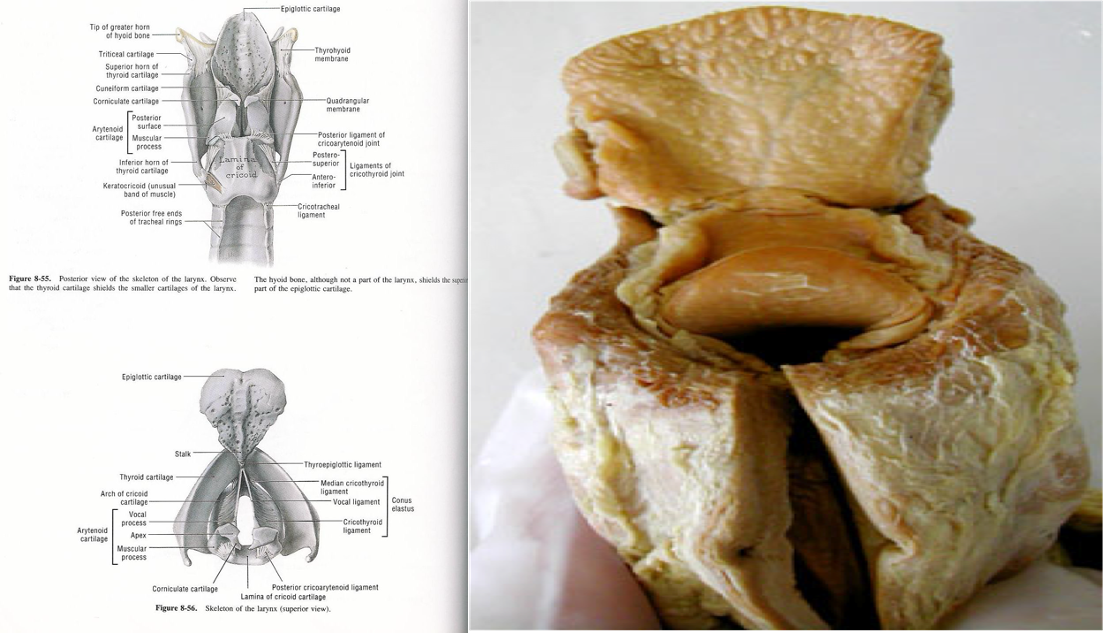
Ligaments
- Thyroepiglottic ligament
- Hyoepiglottic ligament
- Glossoepiglottic fold ► valleculae
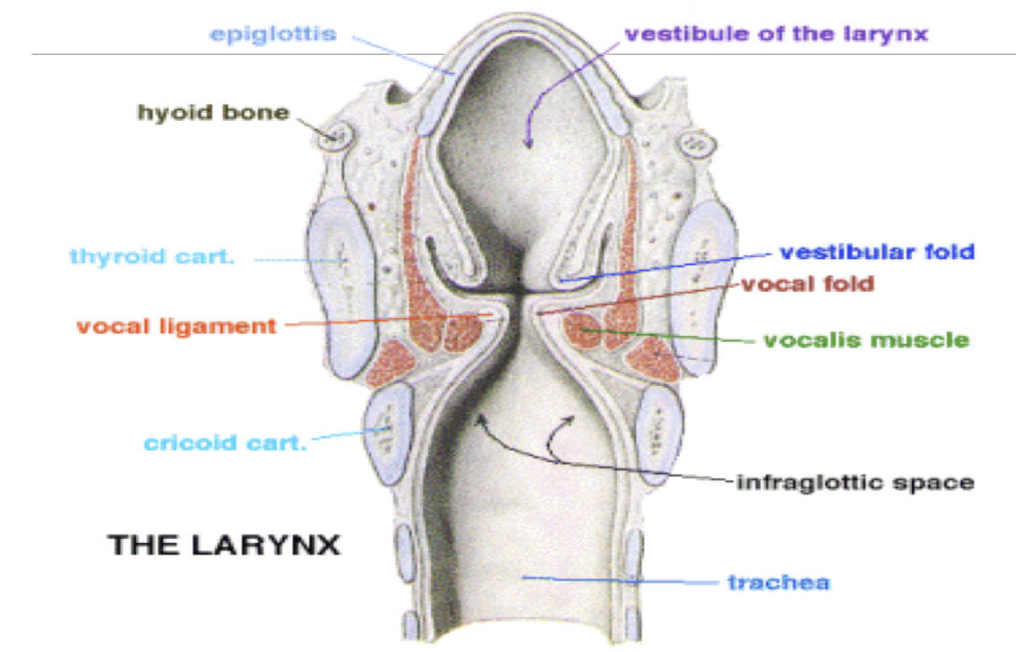
Laryngeal Membranes
Quadrangular Membrane
- Upper and lower border ► thickened
- Aryepiglottic fold
- Vestibular fold
Triangular Membrane (Conus Elasticus)
- Medial and lateral border is free ► thickened ► vocal ligament
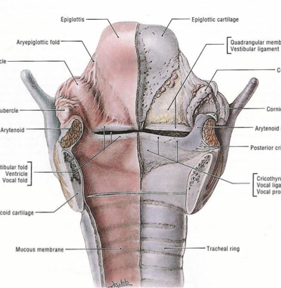
Laryngeal Mucosa
- All mucosa from trachea to aryepiglottic fold ► ciliated columnar epithelium.
- Except vocal cord and aryepiglottic fold ► squamous epithelium
Cavity of Larynx
Laryngeal Musculature
Extrinsic Depressors (C1-C3)
- Sternohyoid
- Sternothyroid
- Thyrohyoid
- Omohyoid
Extrinsic Elevators
- Genohyoid (C1)
- Digastric (CN V, CN VII)
- Mylohyoid (CN V)
- Stylohyoid (CN VII)

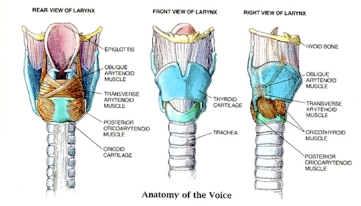
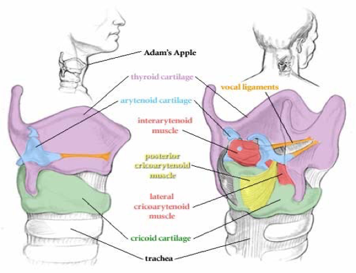
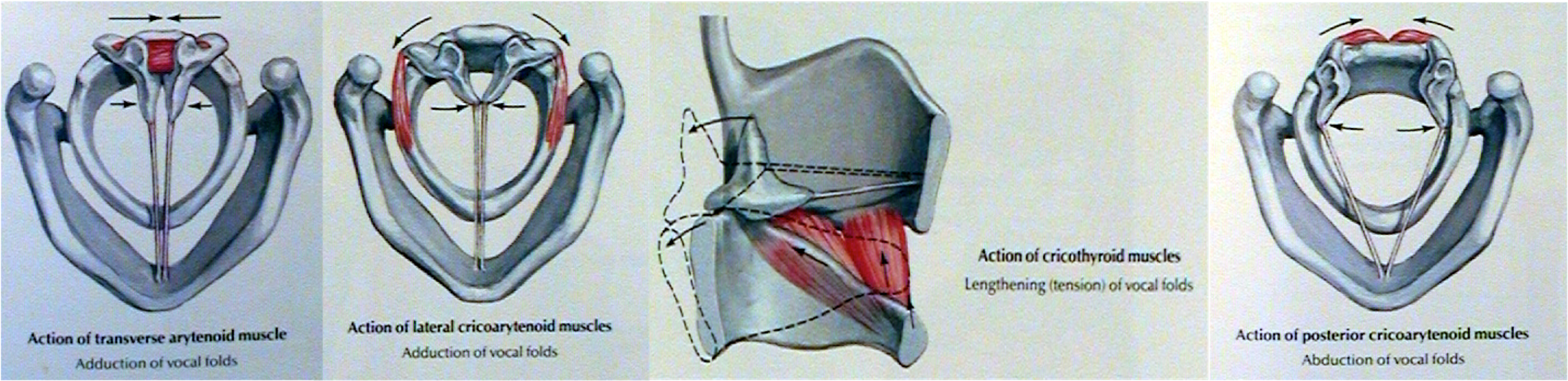
Intrinsic Musculature
Abductors
- Posterior cricoarytenoid (PCA)
Adductors
- Thyroarytenoid (TA)
- Lateral cricoarytenoid (LCA)
- Cricothyroid
- Interarytenoid
Histopathology of the Vocal Cords
Vocal Cord Layers
Histology
- Squamous epithelium
Lamina Propria
- Superficial layer (Reinke’s space)
- Intermediate layer
- Deep layer
- Intermediate + deep layers = vocal ligament
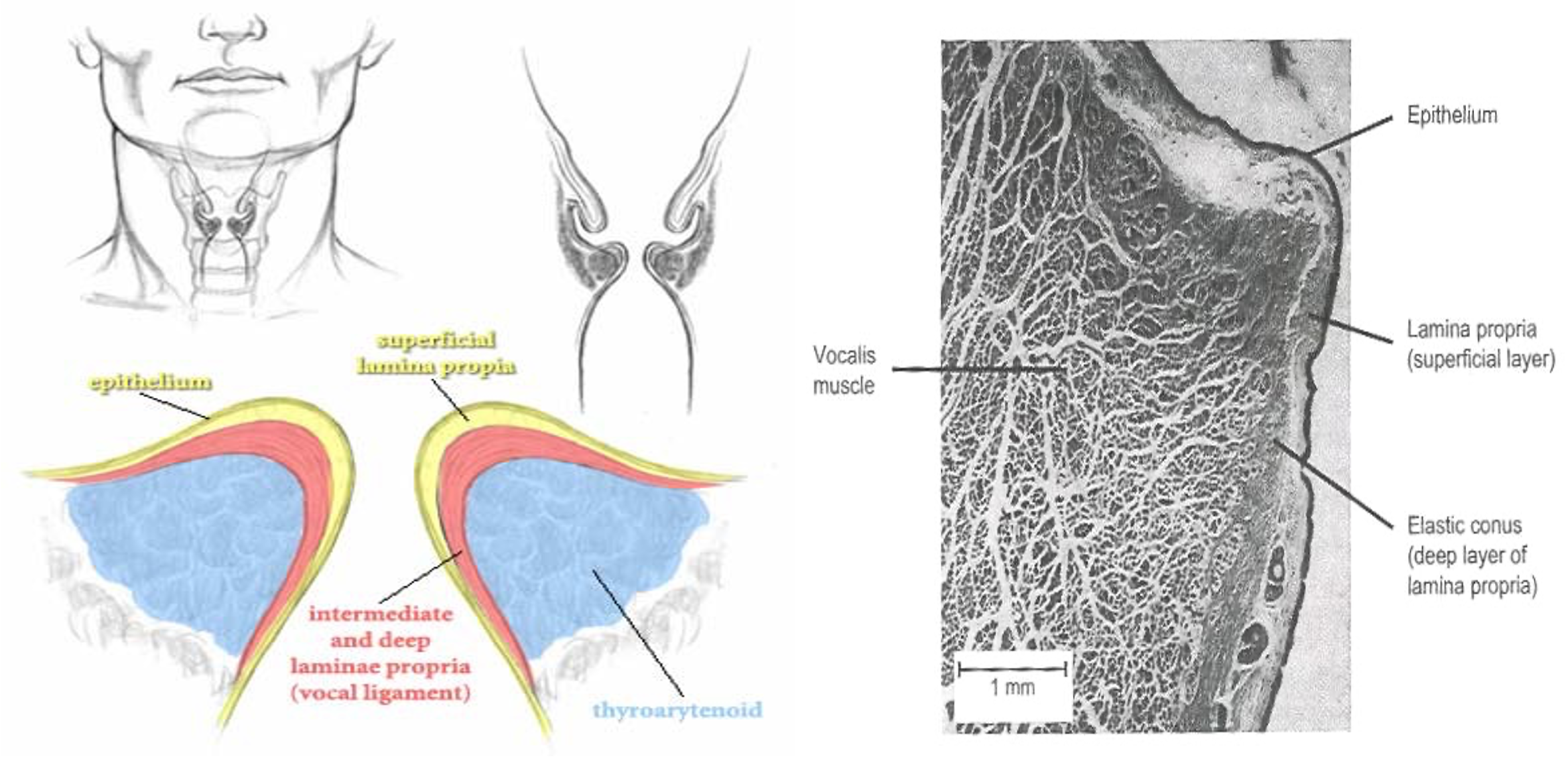
Vocalis (Thyroarytenoid Muscle)
Blood Supply and Lymphatic Drainage of the Larynx
Blood Supply
- Superior and inferior laryngeal artery and veins.
Lymphatic Drainage
- Above vocal cord ► upper deep cervical lymph node.
- Below vocal cord ► lower deep cervical node.
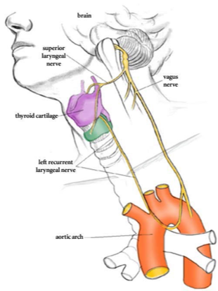
Nerve Supply of the Larynx
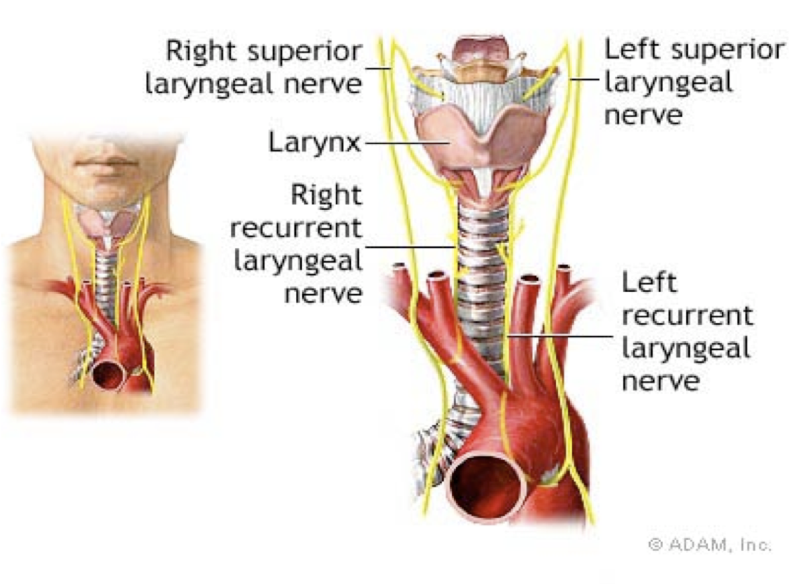
Superior Laryngeal Nerve
- Internal branch (sensory) + superior laryngeal artery.
- External branch ► cricothyroid muscle
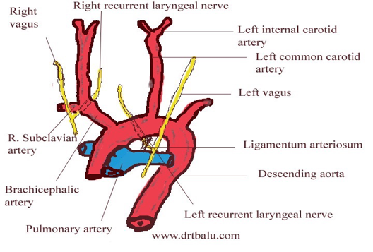
Recurrent Laryngeal Nerve
- Right side: crosses the subclavian artery
- Left side: arises on the arch of the aorta deep to ligamentum arteriosum
- It is divided behind the cricothyroid joint
- Motor ► all the intrinsic muscles except cricothyroid
- Sensory
Pediatric Airway Anatomy
- The neonates are obligate nasal breathers until 2 months.
- The epiglottis at birth is omega (Ώ) shaped.
- The infants have a high larynx (C1-C4).