Internal Medicine
ACUTE PULMONARY EDEMA
Acute worsening of a stable CHF patient can occur due to any of following:
- Non-compliance with diet & meds
- Severe & sudden rise in BP
- Acute MI in a CHF patient
- Sudden arrhythmia, especially atrial fibrillation
- Any infection (e.g., pneumonia)
S/S of acute heart failure/Pulm. edema
There is a sudden onset of pulmonary edema which causes:
- Severe dyspnea & orthopnea
- Restlessness
- Cough with pink frothy sputum
- Pt. looks in severe resp. distress
- Severe crackles & wheezing on auscultation - Lung sounds X
MANAGEMENT
- Propped-up position - 45 degree - relieves shortness of breath Z
- O2
- i.v. Lasix
- i.v. nitrates (e.g., nitroglycerin/nitroprusside). Cause vasodilation which is helpful
- i.v. morphine (reduces restlessness & causes vasodilation)
- i.v. Nesiritide (causes vasodilation & also diuresis)
- Urgent chest X-ray: Will show severe pulmonary edema +/- pleural effusion
- i.v. inotropes (dopamine) if BP is low
Imaging
Pulmonary edema is the accumulation of fluid in the lungs.
cause
- can be cardiogenic (e.g., acute myocardial infarction, congestive heart failure)
- or non-cardiogenic (e.g., pneumonia, blood transfusion, preeclampsia, shock).
Clinical features include:
- Progressive dyspnea
- signs of hypoxemia (e.g., cyanosis, tachycardia).
X-ray chest findings in cardiogenic pulmonary edema
- Central edema
- Kerley B lines: visible horizontal interlobular septa caused by pulmonary edema.
- Pleural effusions.
- Enlarged heart size.
X-ray chest findings in noncardiogenic pulmonary edema
- Patchy and peripheral edema
- Possibly ground-glass opacities and consolidations with air bronchograms
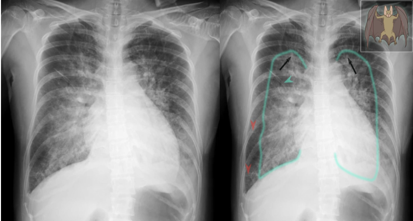
X-ray chest (PA view)
of a patient in the 3rd trimester of pregnancy with peripartum cardiomyopathy
The cardiac silhouette is enlarged and upper lobe vessels are prominent (redistribution of flow). Bilateral interstitial and airspace edema is predominantly perihilar in distribution and produces a classic batwing, or butterfly, appearance. Pulmonary vessel margins are obscured (perihilar haze) and thickened interlobular septae (Kerley lines) are conspicuous.
