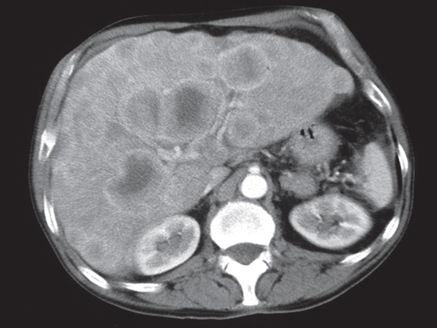
At CT, metastases are seen as rounded areas, usually lower in density than the contrast enhanced surrounding parenchyma, Most are well demarcated from the adjacent parenchyma.
 CT scan of liver metastases
There are a large number of low density lesions in both lobes of the liver, which show enhancement around their edges. The patient had carcinoma of the bronchus.
 Ultrasound of liver metastases.
- (a) Multiple hyperechoic metastases scattered throughout the liver.
- (b) Multiple metastases appearing as well-defined, round, hypoechoic lesions scattered throughout the liver.
- (c) The cursors indicate a metastasis showing reduced echogenicity, but with an echogenic centre known as a target lesion; here is solitary lesion