IM
- An enlarged thyroid gland is called goiter
- Enlargement may be diffuse, partial, smooth, nodular
- Can be associated with S/S of hyperthyroidism or hypothyroidism or even no symptoms (euthyroid state)
ENT
Simple Goiter
Definition
- Non-inflammatory, non-neoplastic, and non-toxic enlargement of the thyroid gland.
Types
A- •Physiological Goitre - diffuse hyperplastic goiter
- It may occur in female at puberty or during pregnancy and lactation.
- It is due to increase body demand, which will lead to relative iodine deficiency
- Patient complains of mild enlargement of the gland
Prevention:
- use iodized table salt (potassium iodide)
Treatment:
- No treatment (mild cases)
- Thyroid hormone is given, L- thyroxin.
2-Colloid Goiter
- Represents a late stage of diffuse hyperplasia.
- The gland is diffusely enlarged, soft, smooth, and symmetrical.
- It may return to normal or cause pressure manifestations.
- Treatment is conservative unless pressure manifestations occur, in which case a total thyroidectomy may be necessary.
B-Simple Nodular Goiter
Incidence
- Female at 30-40 years. Etiology
- Repeated fluctuations of TSH level _ hemorrhage _ necrosis _ fibrosis _ nodule. Symptoms
- Cosmetic deformity (main complaint)
- Pressure symptoms e.g. :Pressure on trachea : positional dyspnea and cough especially at night
- Pain: in hemorrhage or malignancy.
Sign
- Asymmetrical thyroid swelling in lower part of the neck moving up and down with deglutition.
- Nodular firm thyroid swelling.
- Displacement of trachea.
- Shifting of carotid artery pulsation in huge goiter.
lf associated with RSG(Retrosternal G.). there will be:
- Engorgement of neck veins, lower edge can not be reached, dullness over the manubrium sterni.
Complications
1- Pressure on trachea: - Unilateral compression _ kinking of trachea. - Bilateral compression _ anteroposterior slit (scabbard trachea). 2- Secondary thyrotoxic changes (in 30% of cases) 3- Hemorrhage (precipitated by cough or shout): - It is an emergency and treated by urgent aspiration or subtotal thyroidectomy. 4- Cyst formation: very common 5- Infection: very rare. 6- Retrosternal extension. 7- Calcification. 8- Malignant change _ follicular carcinoma (0.5-5%).
Investigations
- Thyroid function tests: normalT3 , T4 ,TSH levels.
- U/S of the neck: to detect cystic or solid.
- FNABC: from dominant nodule to exclude malignancy. To exclude complications: Thyroid scan: to exclude secondary thyrotoxicosis and CT Scan to exclude retrosternal extension.
Preoperative investigations: CBC, KFTs, LFTs, and indirect laryngoscopy.
Treatment
Treatment of SNG
- hemithryoidectomy
- Postoperative L-thyroxine to avoid recurrence in the left thyroid tissue.
Other lines of treatment:
- Total thyroidectomy + replacement therapy
Treatment of complications: toxic goiter:
- Total thyroidectomy after preparation.
- Radio-active iodine.
Malignant changes (Follicular carcinoma):
- Total thyroidectomy+ Supplementary L-thyroxine.
- Radioactive iodine for metastasis.
Retrostemal extension:
- surgical excision.
Hemorrhage:
- Urgent aspiration or urgent subtotal thyroidectomy.
Imaging
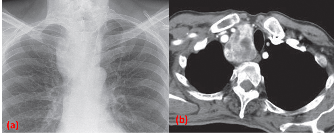
Intrathoracic thyroid masses (goitres):
-
are the most frequent cause of a superior mediastinal mass.
-
The characteristic feature is that the mass extends from the superior mediastinum into the neck and almost invariably compresses or displaces the trachea.
Retrosternal goitre.
(a) X-Ray showing a large, right-sided, superior mediastinal mass displacing the trachea.
(b) CT in the same patient showing the heterogeneously enhancing mass to the right of the trachea
