1) Look:
- Location
- shape
- Size (at least 2 directions
- Floor (Colour & Depth)
- Discharges (pus, blood, quantity)
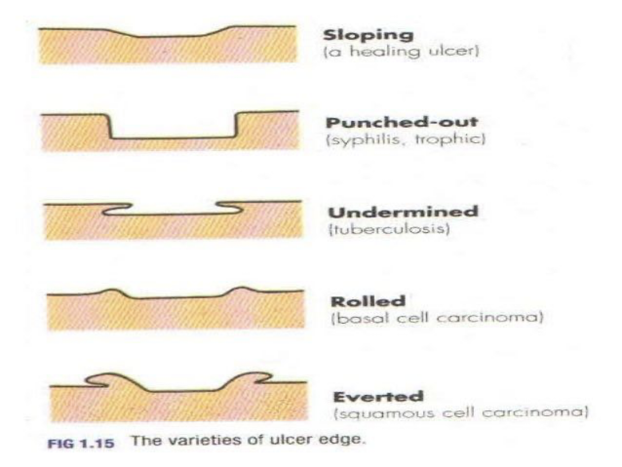
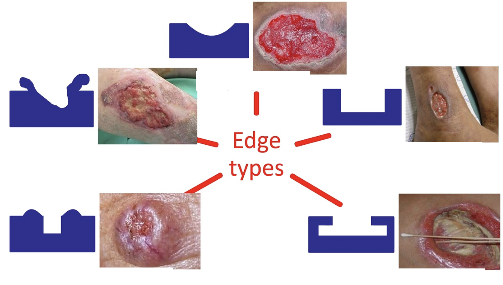
- Edge
2) The Floor: #Z
-
Granulation Tissue (Red Pink): Collagen, fibroblast, capillary, Inflammatory cells and bacteria (during healing process)
-
Eschar: (Black): Layer of dead tissue, become dehydrated and form dark eschar
-
Scab: (yellow): Sloughed and dried small amount of discharges may dry to become scab
(Serous discharge – healing ulcer Purulent discharge – inflamed and spreading ulcer.
Serosanguinous discharge – tubercular ulcer, malignant ulcer. Greenish discharge – infection)
3) Edge: Z
- Flat sloping ( healing / venous)
- Punched out (Syphilis / DM / Ischemic)
- Undermined (pressure necrosis, Carbuncle, Tuberculous)
- Rolled (BCC)
- Everted (SCC / Ulcerated AdenoCA)


4) Feel: on base
- Tenderness
- Temperature
- Base
5) Examine Surrounding Tissue:
- Induration (Infection, Trauma)
- Pigmentation
- Scaring
- Edema
- Vascular Assessment; arterial/venous - Varicose, Ischemia
- Neurosensory Assessment - sensation/movement(flexion:extention)
- Regional Lymph nodes
**Local** (lymphatic / venous obstruction) General (Liver, Cardiac, Renal, low Alb)
