These tumors arise from three different types of cells that are normally found in the brain: astrocytes, oligodendrocytes, and ependymal cells.
-
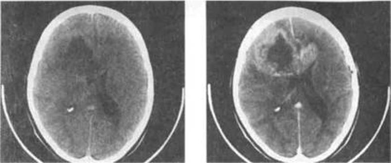
At CT, a glioma typically appears as a solitary, irregular, low attenuation lesion.
-
Local mass effect can usually be demonstrated. Gliomas ***may calcify, particularly ***
oligodendrogliomas.
Glioma on CT pre and post contrast

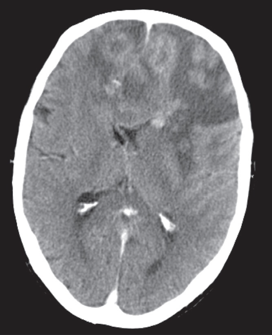
CT scan, post intravenous contrast,
showing an irregular enhancing mass with surrounding low attenuation involving both frontal lobes and the corpus callosum, in keeping with a high grade glioma (glioblastoma multiforme).

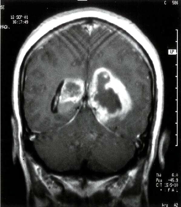
Bilateral glioblastoma (butterfly glioma)
T1-weighted contrast-enhanced MRI: bihemisphere contrast enhancement lesion. There is peripheral contrast agent enhancement. The third ventricle is centrally located. The tumor is displacing the posterior horn of the lateral ventricle .
