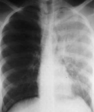
Important Points
-
In atelectasis, there is a shift toward the side of the opacification
-
In pleural effusion, there is a shift away from the side of the opacification
-
In pneumonia, there is no shift
-
In pneumonectomy, the 5th rib is usually absent
Silhouette Sign
The information on a CXR is largely dependent on the contrast between air in the lungs compared with the opacity of the heart, blood vessels, mediastinum and diaphragm.
An intrathoracic lesion touching the heart, aorta or diaphragm obliterates the border of the structure in question. This sign is known as the silhouette sign
-
Silhouette sign: In this case the left border of the heart is not visible.
-
Due: Pneumonia/pleural effusion
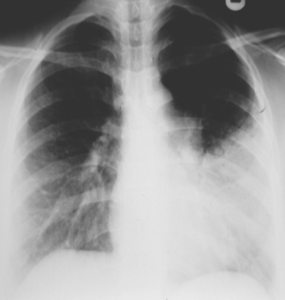
Air-Space opacification
Air-space opacification means replacement of air in alveoli by fluid or other materials (e.g. pus/blood), often called ‘consolidation. signs of** consolidation are:
- • An opacity
- • An air bronchogram
Air-space filling (consolidation) in the right upper lobe is due to pneumonia
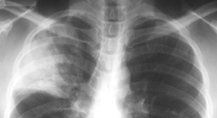
Consolidation of RT. middle lobe in PA and LA view
Collapse consolidation of the middle lobe. Posteroanterior and lateral views. The collapsed lobe is most obvious on the lateral view. Note the silhouette sign obliterating the lower right heart border.
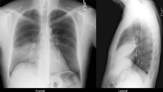 Lateral: Arch of heart - oblique/Horizontal fissures
Abnormal Opaque & Translucency
Opaque: CONSOLIDATION/Pneumonia, Pleural Effusions, COLLSPSE, AGENESIS, PNEUMONECTOMY
TRANSLUCENT:
Emphysema, Pneumothorax, MASTECTOMY, Technical Rotation
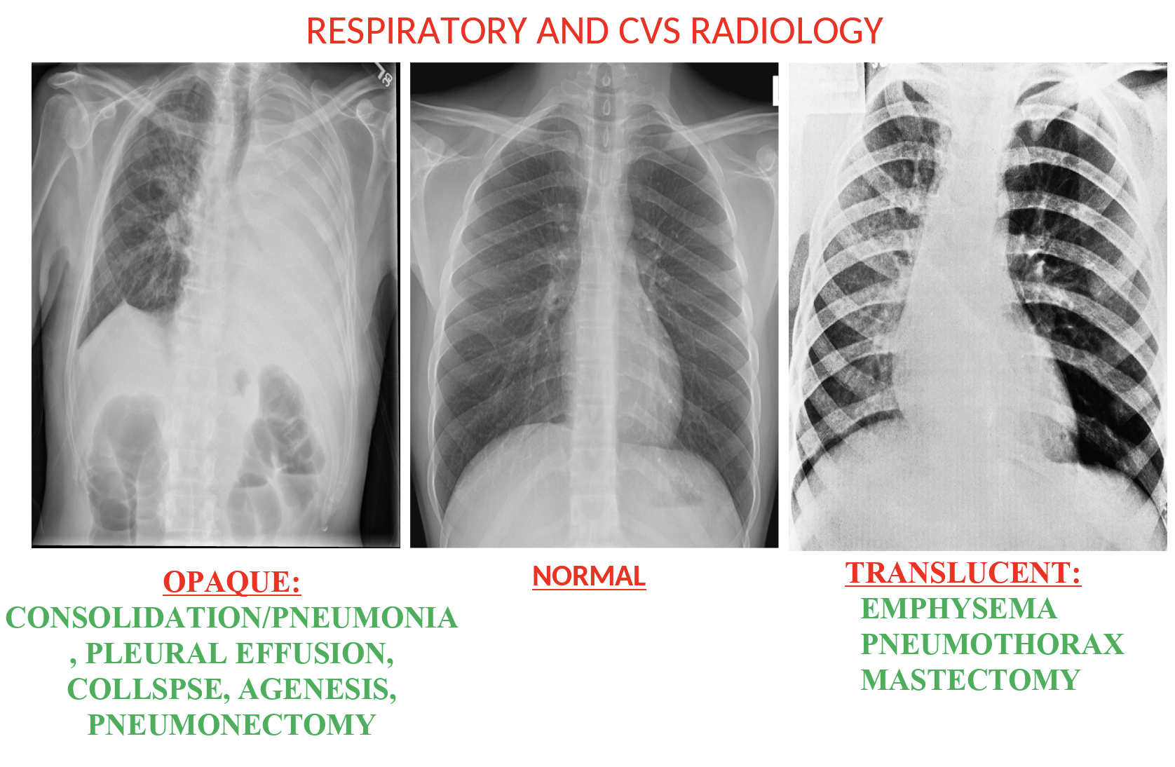
The left hemithorax is opaque
- There is no shift of the heart or trachea
- The opacified hemithorax contains air bronchogram
- No loss of lung volume
