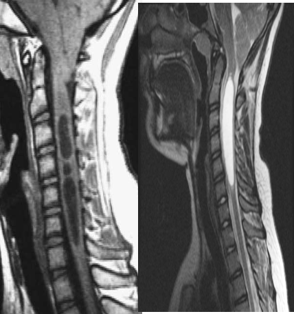
is an abnormal fluid-filled dilation of the central canal of the spinal cord occurring as a result of impaired CSF flow.
Imaging shows focal or diffuse cord expansion.
CT Scan:
Show distinct area of low density within the cord without enhancement at the post-contrast study.
MRI:
Cystic area within the cord which appears of low signal at the T1WI and of high signal at the T2WI. No enhancement seen within the lesion at post-contrast study.
