Pituitary tumors are divided into :
- Macroadenomas (>1 cm)
- Microadenomas (<1 cm).
Imaging studies
MRI sella with IV contrast (gold standard)
Indications
- First-line diagnostic modality for suspected pituitary adenomas
- Postsurgical surveillance after resection of a pituitary mass.
Characteristic finding:
Intrasellar mass
Potential additional findings
- Compression of adjacent structures (e.g., impingement on the optic chiasm).
- Hemorrhage and necrosis
- Cavernous sinus invasion
CT sella with IV contrast
Indications
- Second-line diagnostic modality
- Can be used to plan transsphenoidal hypophysectomy
Supportive findings: similar to MRI
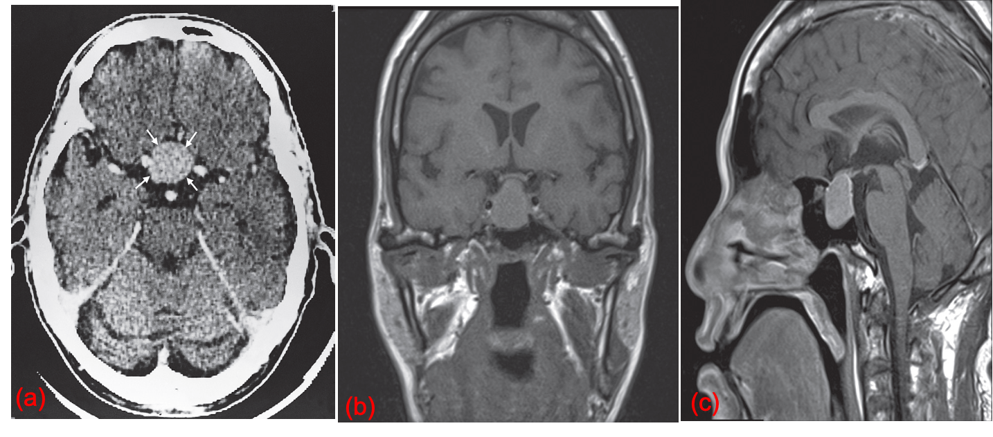
 Pituitary tumor. (a) CT scan after contrast showing a mass in the pituitary fossa which enhances vividly (small arrows).
(b) Coronal T1 MRI and (c) Sagittal T1 post contrast MRI showing a macroadenoma expanding the pituitary fossa and extending superiorly to touch the optic chiasm in the suprasellar cistern.