Retinal Vascular Occlusion
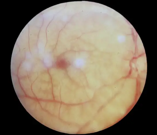
Retinal Vein Occlusion
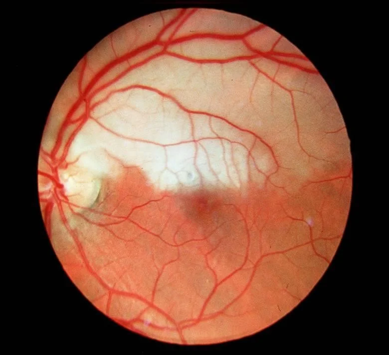
- Ophthalmoscopes picture of disc swelling, venous engorgement, cotton wool spots, and diffuse retinal hemorrhages like blood and thunder.
- Loss of vision may be severe.
- no pain
- There is no generally accepted acute management. Central retinal vein occlusion is not a true ophthalmic emergency.
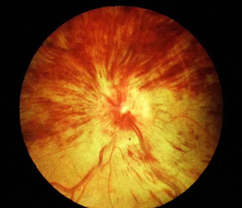
Central Retinal Vein Occlusion
-
***pain de *** iris
-
New Vasculatration of Iris
-
No Pain
-
Severe loss of vision
-
After 3 months Severe Pain and
-
More loss of vision
-
Neovascularization (NV) of Iris
- RI
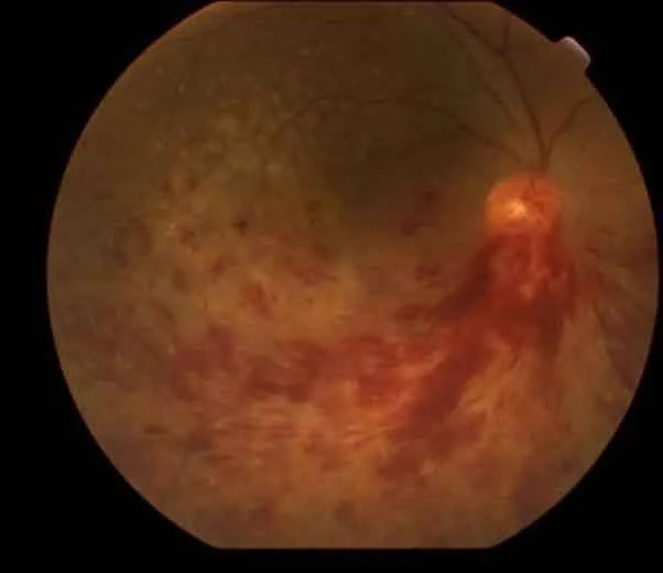
Inferior Macular Lesion
- Inferior
- Macular lesion
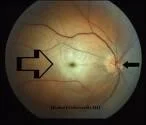
Central Retinal Artery Occlusion
A sudden, painless, and often complete visual loss may indicate central retinal artery occlusion.
Several hours after a central retinal artery occlusion, the inner layer of the retina becomes opalescent.
A cherry red spot is seen due to the pallor of the perifoveal retina in contrast to the normal color of the fovea.
A chronic cherry red spot is also a feature of the storage diseases such as Tay-Sachs, Pick disease, and Niemann-Pick disease.

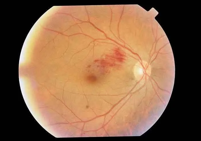
Branch Retinal Artery Occlusion
When only a branch of the central retinal artery is occluded, vision is only partially lost. This is more likely to be the result of an emboli and the source of the emboli should be sought. Cardiologist حواء
If the visual acuity is affected, attempts should be made to dislodge the emboli by ocular massage.
Hemi-Retinal Artery Occlusion