The classic clinical manifestation
of EDH is an initial loss of consciousness, followed by a lucid interval in which the patient gains normal or near-normal consciousness, followed by rapid neurological decline.
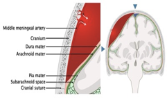
- 90% is arterial (middle meningeal artery)
- Biconvex, lens-shaped
- Commonly associated with skull fractures.
- CT head without IV contrast is the first line imaging exam

EDH Imaging
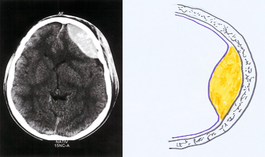
Characteristic findings Biconvex (lenticular shaped), sharply demarcated extraaxial lesion.
- Typically hyperdense in appearance
- Limited by suture lines
- Evidence of skull fracture, if present
Epidural hematoma- CT head (axial section; noncontrast)
An epidural hematoma is visible as a solitary biconvex hyperdense lesion in the left frontotemporal region, causing a midline shift to the contralateral side.

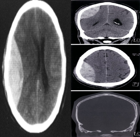
Cranial vault fracture with epidural hematoma CT head (without IV contrast; top: brain window, coronal plane; middle: brain window, axial plane; bottom: bone window, axial plane)
An epidural hematoma (EDH) is visible as a biconvex, heterogeneous, hyperdense extra-axial collection in the right hemicranium. Mass effect on the adjacent cerebral parenchyma and right lateral ventricle has produced contralateral midline shift . The hematoma does not cross the coronal or the lambdoid suture. A scalp hematoma is present, and a fracture of the underlying right parietal bone is visible on the bone window image.
