Layers of the Skin
-
Layers of the Skin
- Epidermis: layers, cell types, and functions
- Dermis: cell types, and functions
- Subcutis
-
Annexal Structures
-
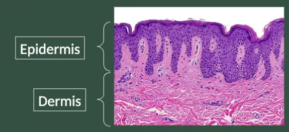
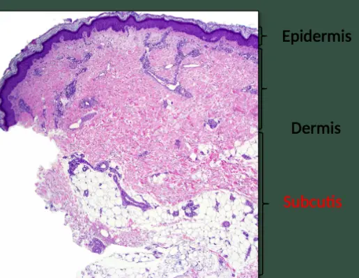
The skin is composed of three layers:
- Epidermis
- Dermis
- Subcutis
Layers of the Skin
- Epidermis; The epidermis is the topmost layer, and consists primarily of keratinocytes.
- Dermis; lies below the epidermis, and consists primarily of fibroblasts, collagen, and elastic fibers.
- The dermis lies below the epidermis and consists primarily of fibroblasts, collagen, and elastic fibers.
- Below the dermis is also called subcutis, panniculus, or hypodermis.
Outline: Basic Anatomy of the Skin
- Layers of the Skin
- Epidermis: layers, cell types, and function
- Dermis: layers, cell types, and function
- Subcutis
- Adnexal Structures
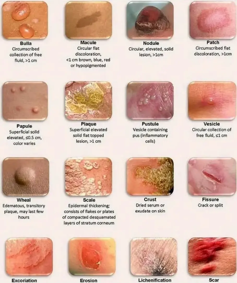
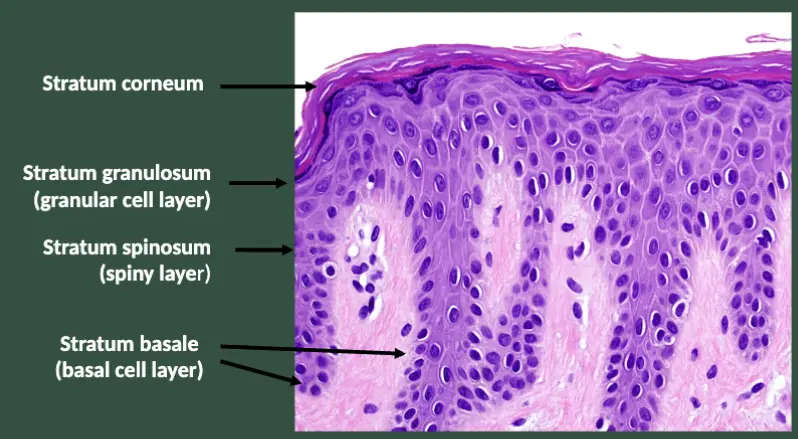
Can you name the four major layers of the epidermis?
-
Stratum Corneum
- This is the outermost layer of the epidermis. It consists of dead, keratinized cells that are constantly shed and replaced in 14 days cycle.
-
Stratum Lucidum
- Found only in thick skin (like the palms of the hands and soles of the feet), this layer is composed of clear, flat, dead keratinocytes.
-
Stratum Granulosum
- This layer contains cells that are undergoing apoptosis (programmed cell death) and are filled with keratin.
-
Stratum Spinosum
- Also known as the prickle cell layer, it is composed of several layers of keratinocytes that are connected by desmosomes.
Functions of the Epidermis
Epidermal cells mature and differentiate over their two-week life cycle from the basal layer to the stratum corneum. They are then shed two weeks after reaching the stratum corneum (for a 28-day cycle).
-
Basal Layer
- The basal layer is the source of epidermal stem cells.
- Cell division occurs here.
- Keratinocytes start in the basal layer and move upwards.
-
Spinous Layer
- Center of epidermis.
- Has a “spiny” appearance due to desmosomal junctions which hold the keratinocytes together.
-
Granular Cell Layer
- Lipids produced by the keratinocytes in the granular cell layer and secreted into the extracellular space between the keratinocytes form a water barrier that keeps water in the skin.
-
Stratum Corneum
- Made up of desquamating keratinocytes.
- Thick outer layer of flattened keratinized non-nucleated cells (corneocytes) provide a barrier against trauma and infection.
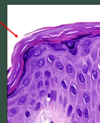
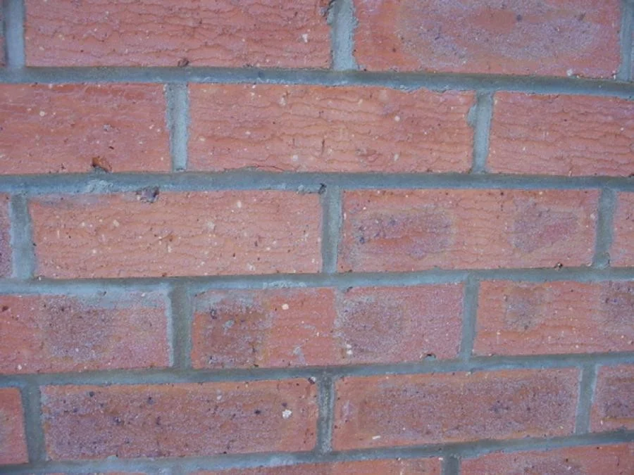
The Stratum Corneum
- You can think of the stratum corneum as a wall of bricks and mortar:
- Bricks: flattened keratinocytes filled with keratin and filaggrin
- Mortar: lipid mixture, which surrounds the keratinocytes and provides the water barrier
Diseases Related to Dysfunction of the Epidermal Layers
- Certain diseases cause loss of adhesion:
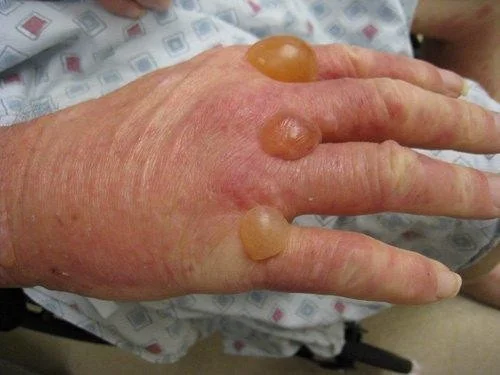
- Bullous Pemphigoid: an autoimmune blistering disease, typically affects older patients.
- Autoantibodies form to antigens directly beneath the basal layer of the epidermis.
- Clinically, presents as tense bullae on an erythematous base on the skin.
Blister Analysis (Bullous Pemphigoid)
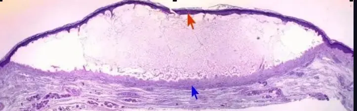
- The epidermis forms the roof of the blister.
- The dermis forms the base of the blister
Diseases Related to Dysfunction of the Epidermal Layer
- In psoriasis, the rate of epidermal turnover is increased (thickening).
- The accelerated rate of movement through the epidermis doesn’t allow adequate time for differentiation, which is recognized as scale.
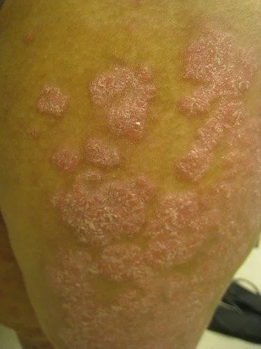 Erythematous, well demarcated
Plaque = raised
patch = color
scaling, redness
Erythematous, well demarcated
Plaque = raised
patch = color
scaling, redness
Accumulation hyper production of cells cycle for 3 days.
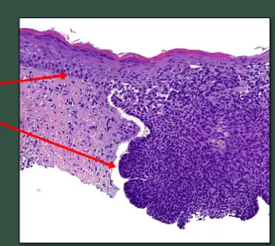
Can you name the type of skin cancer?
See if you can identify this common skin cancer based on what you have learned about the layers of the skin. Hint: The cells composing this growth resemble what layer of the epidermis?
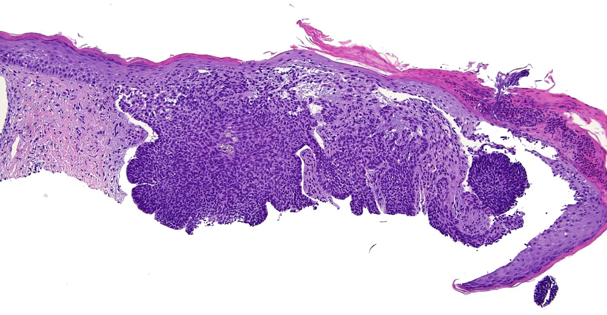
Basal Cell Carcinoma
- Most common form of skin cancer.
- Composed of cells that resemble basal keratinocytes.
Basal Keratinocytes