Landmarks of the Neck
Surface Landmarks
- Hyoid bone
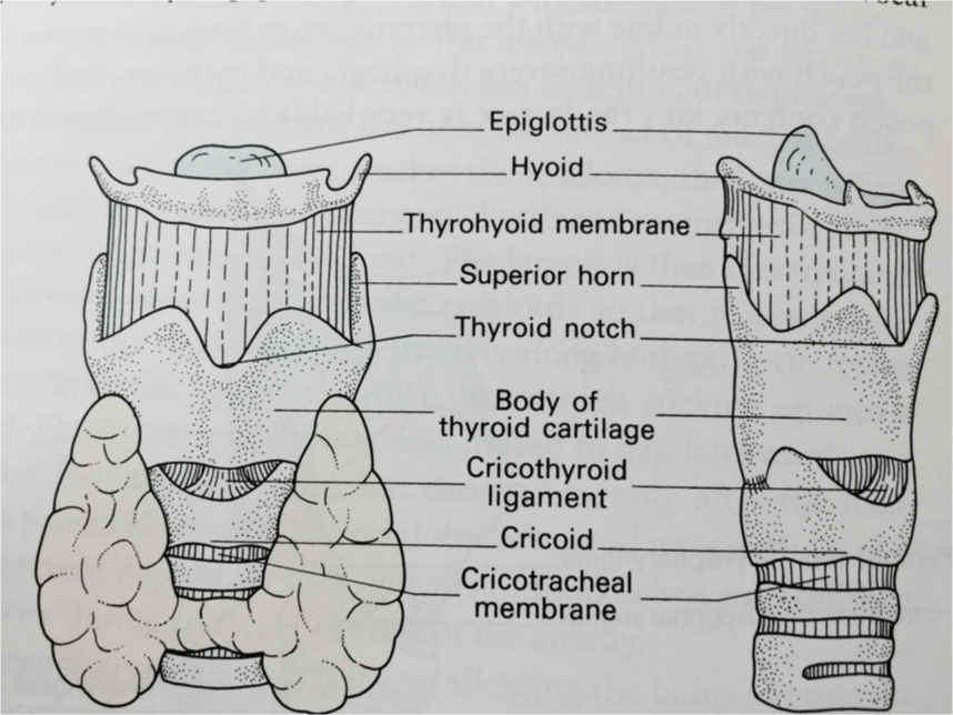
- Laryngeal prominence
- Arch of cricoid cartilage
- Sternocleidomastoid
- Greater supraclavicular fossa
- Trachea
- Suprasternal fossa
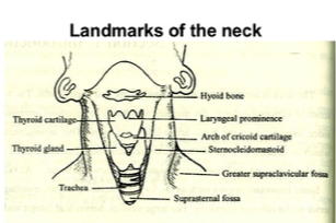 Fig. 2-2 Surface landmarks of the neck (The head is fully extended)
Neck Triangles
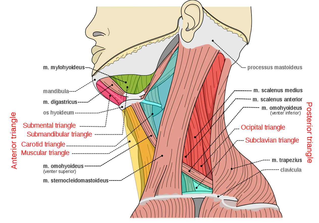
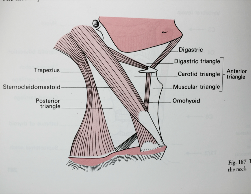
The sternocleidomastoid muscle divides each side of the neck into two major triangles:
-
Anterior Triangle
- Submandibular triangle
- Submental triangle
- Carotid triangle
- Muscular triangle
-
Posterior Triangle
- Occipital triangle
- Supraclavicular triangle
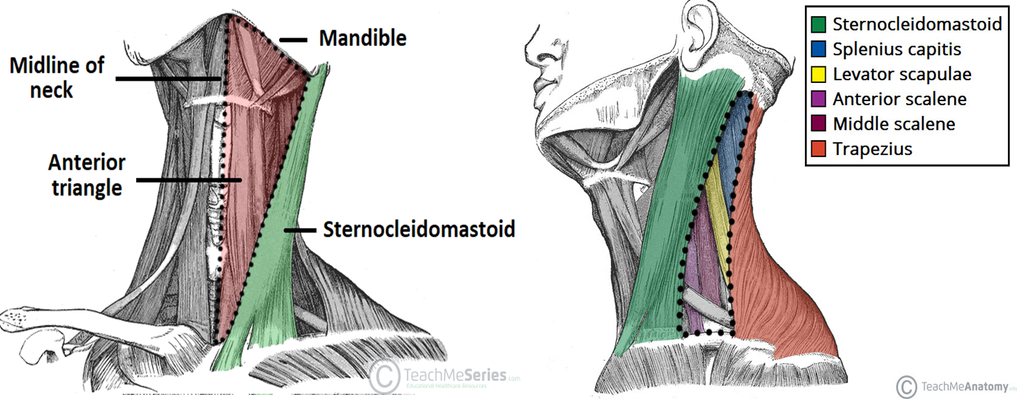
Anterior triangle Borders: - Laterally: posterior border of the SCM - Medially: midline - Superiorly: lower border of the mandible Posterior triangle Borders: - Anteriorly: posterior border of the SCM - Inferiorly: clavicle - Posteriorly: anterior border of trapezius muscle
Anatomy:
The anterior triangle is situated at the front of the neck.
It is bounded:
- Superiorly Inferior border of the mandible (jawbone).
- Laterally Anterior border of the sternocleidomastoid.
- Medially Sagittal line down the midline of the neck from the chin to the manubrium .
The posterior triangle is situated posterior to SCM
Its boundaries are as follows:
- Anterior Posterior border of the SCM.
- Posterior Anterior border of the trapezius muscle.
- Inferior Middle 1/3 of the clavicle.
SURGE Z
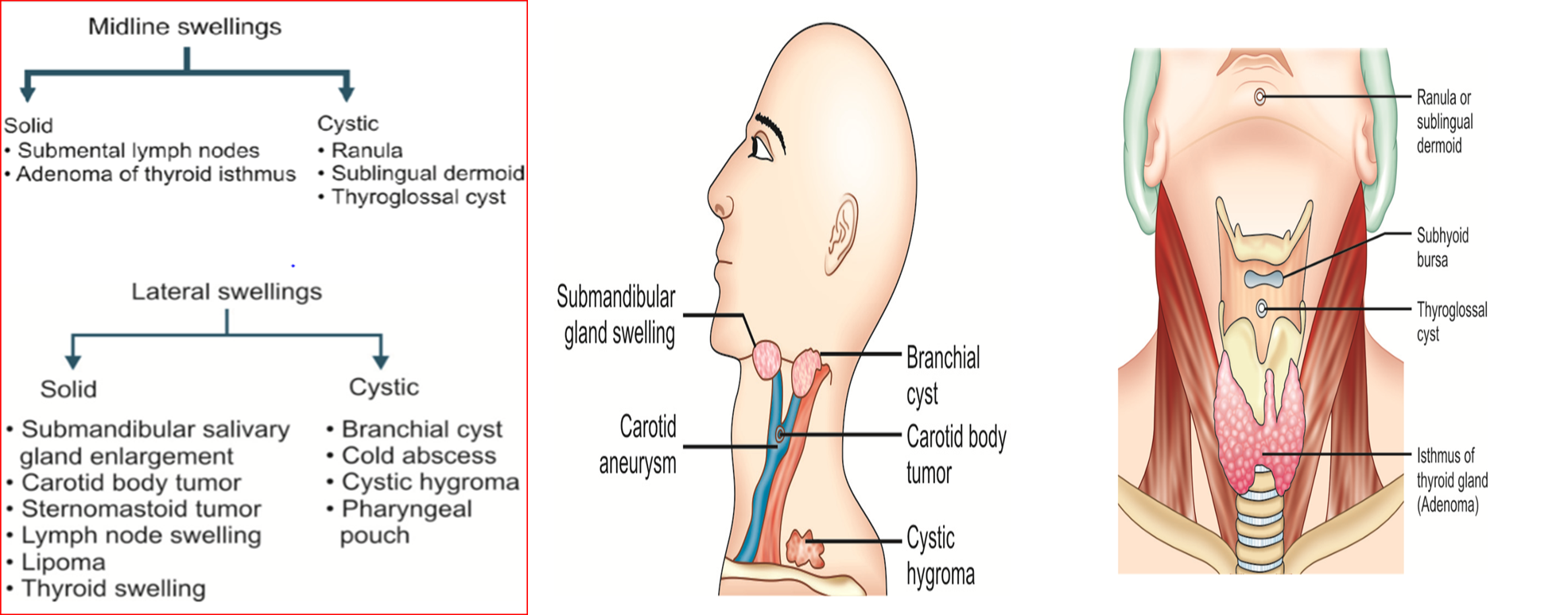
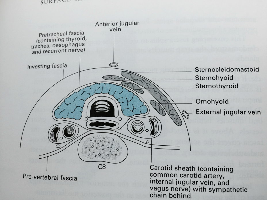
- Midline limited by sternocleidomastoid
- 3rd img at level of C8
- Reccurent laryngeal nerve
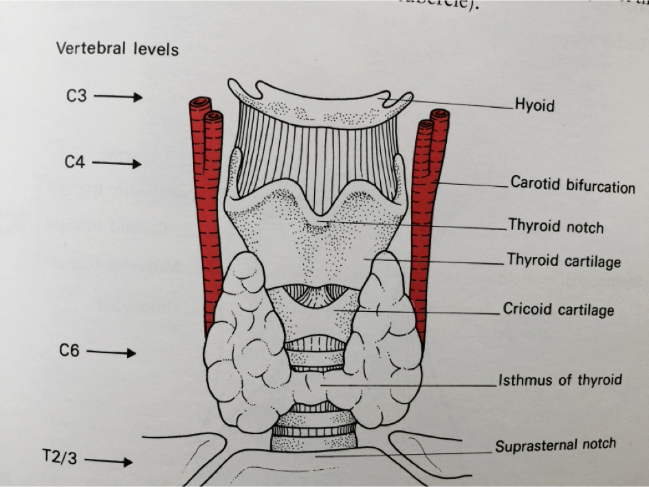
- carotid
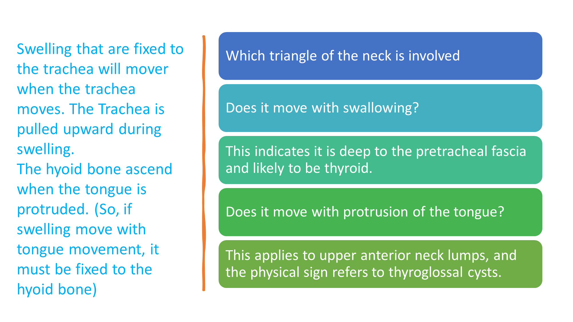
- Larynx goes up then forward - if swelling goes up and down - thyroid
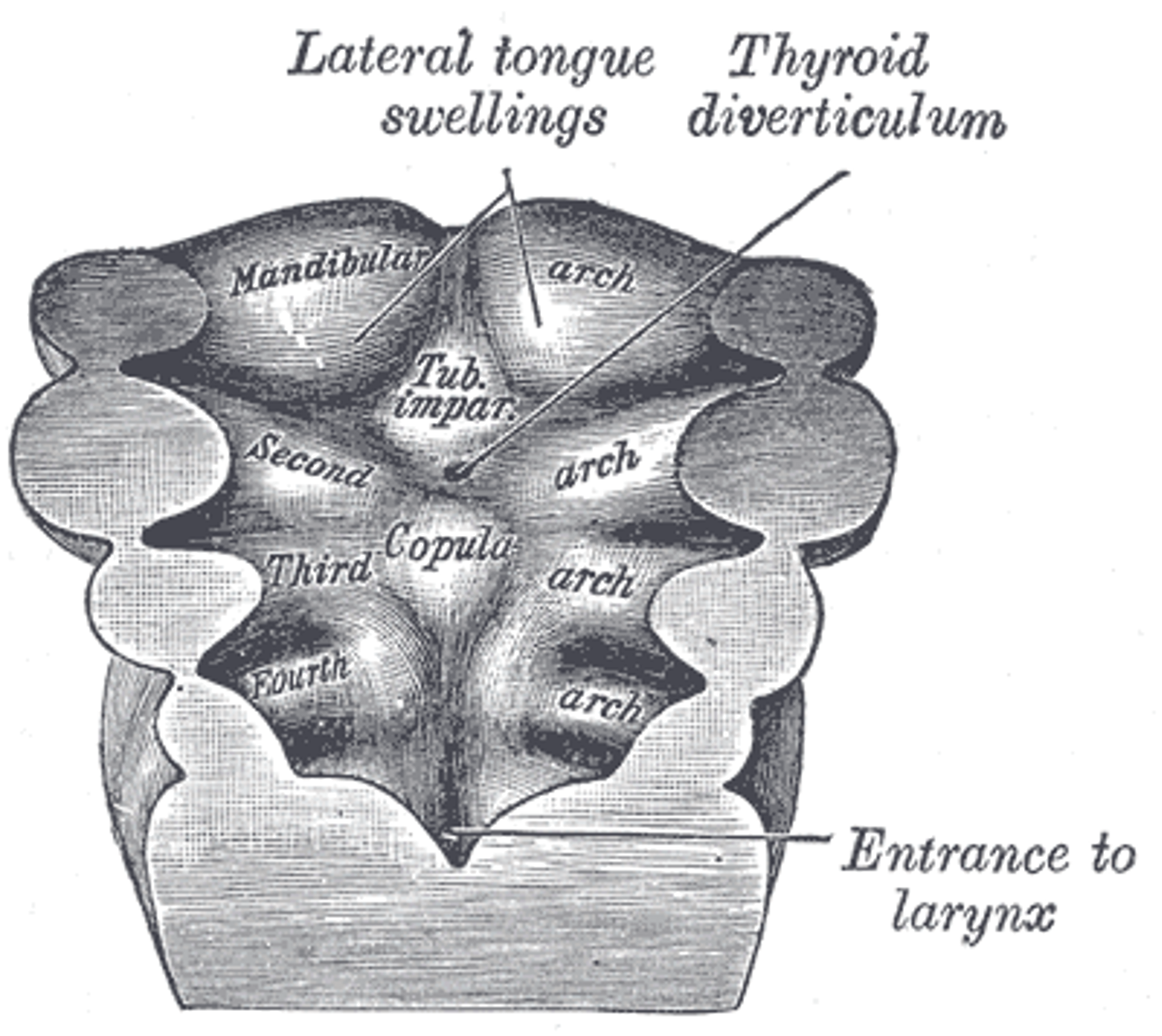
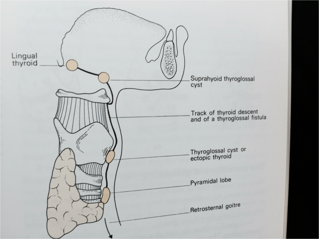 cyst
cyst
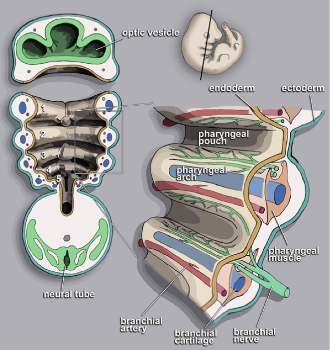 branchial cyst
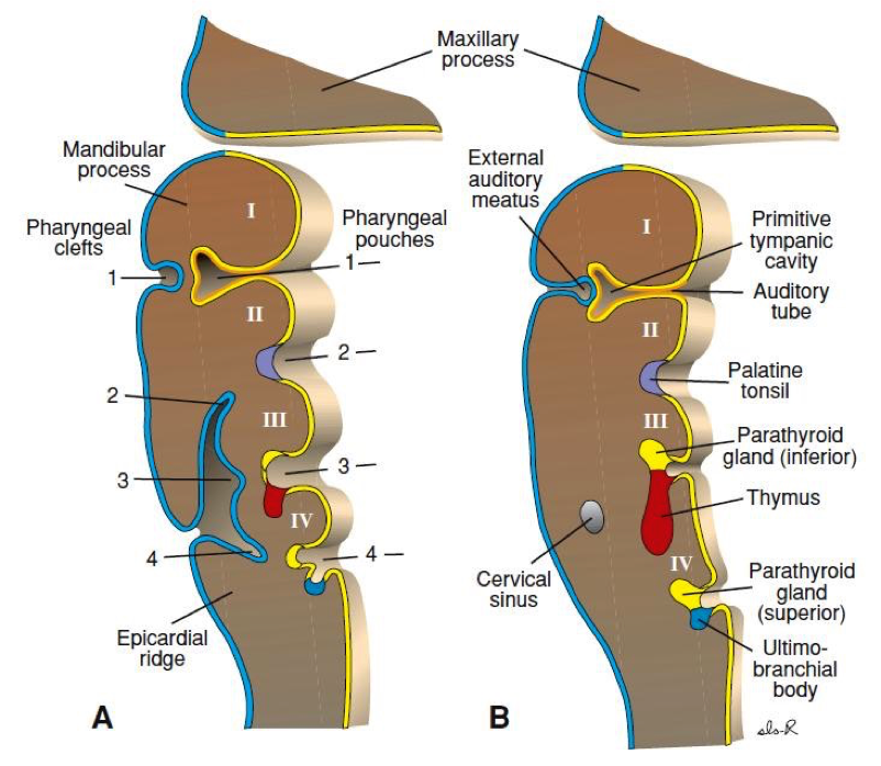
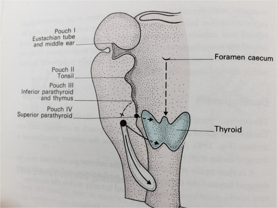
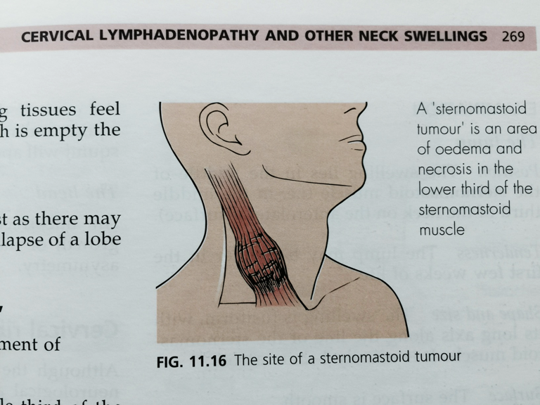
torti - — rhabdomyosarcoma extremely rare
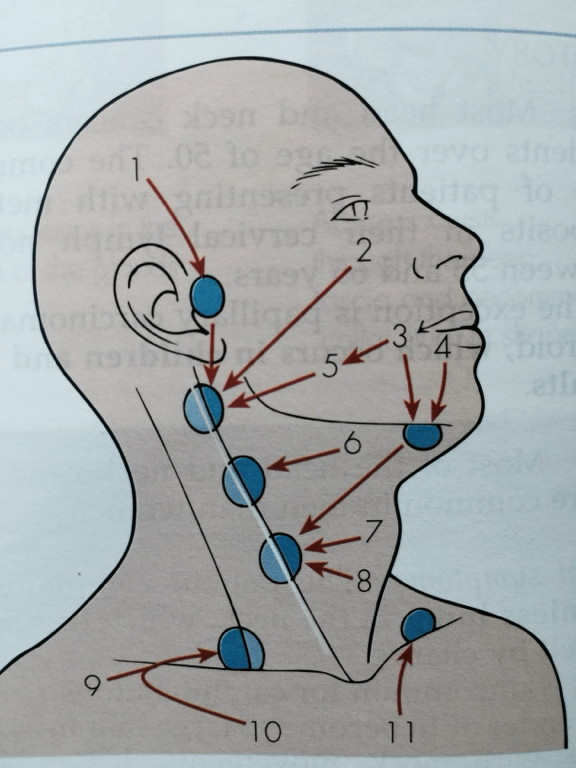 mutliple = lymphonadopathy
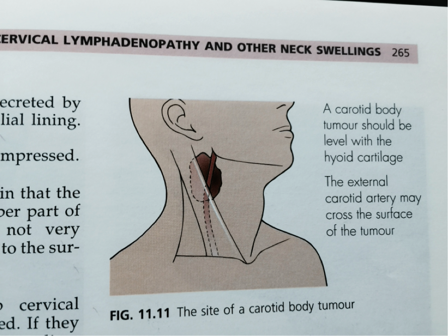 carotid body tumour - glomerulus tumour, pituatiory …
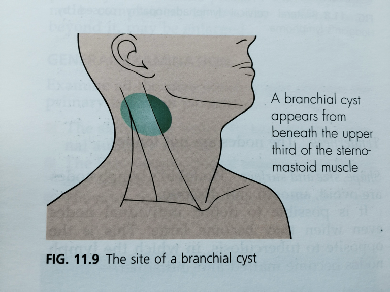 anterior border - may rupture
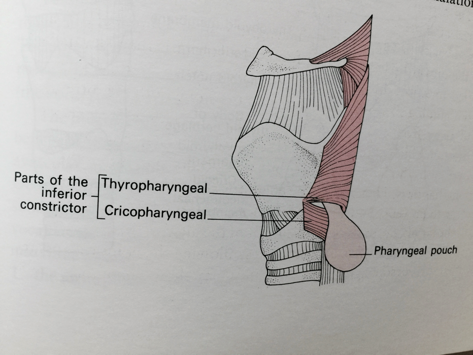 False diverticulum