The importance of general examination:
- Helps to determine the most body system should be stressed during systems examination.
- With proper history the general examination may be enough to obtain the diagnosis OR a short list of diagnoses.
A. General Examination Components
- General patient condition
- Face
- Fever
- Pallor
- Jaundice
- Cyanosis
- Lymph nodes enlargement
- Hands, Digits & Nails
- Mouth, Oral cavity & Tongue
- Temperature
- Pulse rate
- Blood pressure
- Respiratory rate
1. The general condition of the patient
- Well
- unwell
- ill
- In pain
- cachexic
- Consciousness level
- delirium
- orientation
2. Face
- A specific diagnosis can sometimes be made by inspecting the face
- Acromegalic
- Down Syndrome
- Cushingoid
- Parkinsonian
2. Fever
- High grade fever
- In history if it is there should be analyzed regarding its course (continuous, intermittent, remittent), association (rigors, sweating), timing (nocturnal), reliving (antipyretic drugs)
3. Pallor
- Indicates anemia (low hemoglobin concentration)
- Skin, mucous membranes
4. Jaundice
- Yellowish discoloration of the sclera of the eyes, mucous membrane, and skin
- Types of jaundice ?
- Prehepatic
- Hepatic
- Post hepatic
- If it is associated with pallor → indicates prehepatic
- if it is associated with pruritus → indicates post hepatic
- If it is associated with fever → may indicates hepatic (viral hepatitis )
5- Cyanosis
- Bluish discoloration of the extremities and tongue
- If more than 50 g/L of deoxygenated hemoglobin is present in the capillary blood, the skin will have a bluish tinge.
- If the tongue is involved → central (cardiopulmonary) cyanosis
- If the digits only involved → peripheral (vasoconstriction) cyanosis
- Raynaud’s phenomena → connective tissue diseases
- Can cyanosis and pallor concomitant together in the same patient?
6- Lymphadenopathy
Complete examination of all LN groups
- Localized
- Generalize
- Discrete or Matted
- Tender
- If it is generalized and associated with pallor may indicates lymphoma
- If it is tender may indicates infections
7- Hands, Digits and Nails
- Tremors
- Fine of stretched hands→ hyperthyroidism
- Rest tremors → Parkinsonism
- Intention tremors→ cerebellar ataxia
- Sweating and erythema → hyperthyroidism
-
Digits bluish discoloration and ulcers→ Raynaud’s phenomenon as part of scleroderma
-
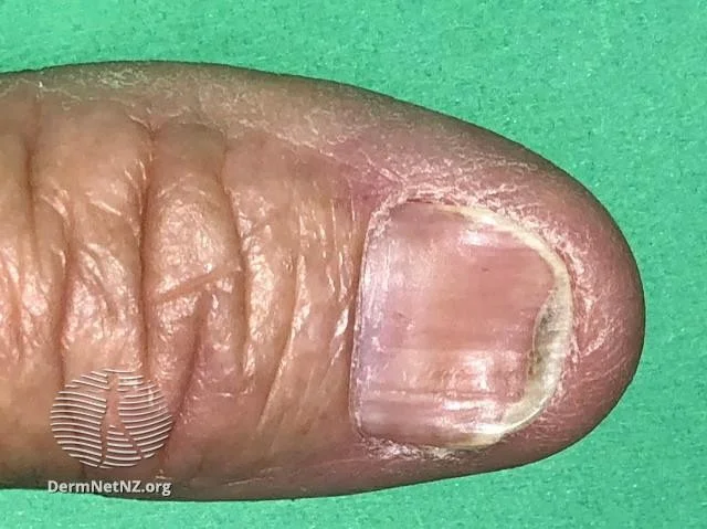
Nail changes may indicate dermatological or systemic diseases
-
Dermatological changes might be due nail infection as onycholysis or a part of generalized dermatological diseases as psoriasis or lichen planus
-
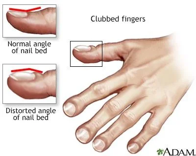
Clubbing fingers → chronic diseases (pulmonary , hepatic ,cardiac, )
koilonychia
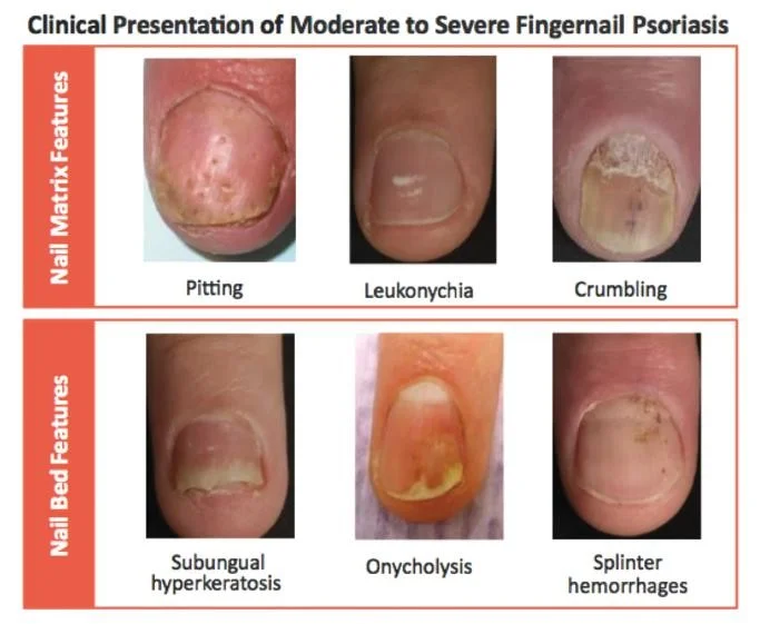
Clinical Presentation of Moderate to Severe Fingernail Psoriasis
Nail Matrix Features
- Pitting
- Leukonychia
- Crumbling
Nail Bed Features
- Subungual hyperkeratosis
- Onycholysis
- Splinter hemorrhages
Clubbed fingers
10- Mouth, Oral cavity & tongue
- Angular cheilitis → anemia
- Smooth tongue →
- Hairy tongue→
- Deviation of the tongue→
- Ulcers →
- Ulceration of the mucous membranes→
- Dental cavities →
- Leukoplakia →
Angular cheilitis
13. Temperature
Measure the core temperature using thermometers
- Digital
- Mercury Sites
- Mouth → 36.8 c
- Axilla → 36. 4 c
- Rectum → 37.3 c
14. Pulse Rate
-
Peripheral arteries ➤ Carotid arteries ➤ Upper extremities→ radial , brachial , ➤ Lower extremities → femoral , popliteal , dorsalis pedis ,
-
Use the index and middle finger , don’t use thumb?
-
Rate → normal , tachy or bradycardia
-
Regular or irregular
-
Detect the character of pulse is it weak thready ,strong , synchronize , radio femoral delay
-
Collapsing pulse → hyperdynamic circulations (anemia , pregnancy , thyrotoxicosis )
15- Blood Pressure
- Using the symphmomanometer (digital or mercury)
- Palpation method → systolic pressure
- It is better to use the two method simultaneously start by palpation method
- Pulse pressure → systolic pressure - diastolic pressure < 60 mmHg
16. Respiratory rate
- Count it for full minute
- Count it while you are pretending takink pulse rate to reduce patient stress ?
- Comment is it normal
- Shallow , deep , apneao , regular or irregular
17. Neck pulsation
- Arterial
- Venous
How we can differentiate?
B. Systems Examination
- The four gold standards of clinical examinations are:
- Inspection
- Palpation
- Percussion
- Auscultation
- The importance and application of each standard differs from one system to other
The Importance of the Four Examination Standards in Different Systems
- General Examination Inspection Palpation
- CVS Auscultation Inspection Palpation Percussion
- Res Auscultation Percussion Palpation Inspection
- GIT Palpation Percussion Inspection Auscultation
- Muscle Inspection Palpation
- Integumentary system Inspection Palpation
- CNS Inspection Palpation
The Skin Exam
- The Total Body Skin Exam (TBSE) includes inspection of the entire skin
- surface, including:
- the scalp, hair, and nails
- the mucous membranes of the mouth, eyes, anus, and genitals
TBSE
- Do not forget the so-called “hidden areas” - places on the skin where lesions may be easily missed
- Conchal bowl (concavity adjacent to the external auditory meatus), auditory canal, postauricular creases
- Medial canthi (angular junction of the eyelids), alar (nasal) grooves
- Intergluteal cleft and perianal skin
- Interdigital spaces
Indications for a TBSE
1- Personal history of skin cancer
2- Increased risk for melanoma I-Two first-degree relatives with melanoma II- Over 100 nevi (moles)
3- Patient with concerning or changing growth
4- New rash on body
5- New patient with undiagnosed skin condition
6- Follow-up patients with extensive skin conditions such as psoriasis
Essential elements for the skin exam
- Adequate lighting
- Undressed patient, in a gown - Preferably without makeup, watches, jewelry
- Privacy
- Ruler
- Magnifying glass
- An open mind about what you are seeing
Getting started
1- Lighting
- The skin exam should be performed with adequate lighting
- natural sunlight is best
- if windows are in the exam room, open the blinds
- the best artificial source is high-intensity incandescent light
- If lighting is too low, turn on as many lights as possible and position the patient directly under available lights
2- Undressed patient
- You cannot diagnose what you cannot see
- Before starting the skin exam, ask the patient to undress to their bra and underwear and put on a gown with the opening to the back
- Put down a chux or exam table paper so their bare feet don’t touch the floor
- Tell the patient you will step out, and ask if they would like a chaperone during the exam
- If you expect to examine the breasts or genitalia of an opposite-gender patient, bring a chaperone regardless
- Draw the curtain and step out of the room
3- Patient modesty
- Undressed patients feel very vulnerable
- Avoid keeping them waiting too long while undressed
- Offer a second gown or blanket if it is cold
- Before untying a gown or moving it, ask permission
- Ask the patient to expose the area being examined, and cover the area after it has been examined
- Say out loud what part of the body you want to examine next
- e.g., “Okay, now let’s look at your chest and abdomen”
- The patient will usually move the gown accordingly
4- Sanitize your hands
- The skin exam is tactile as well as visual
- You must palpate lesions to tell if they are raised, flat, or atrophic
- Many dermatologists prefer to use gloves for moist areas (groin, axilla) or oozing, crusted lesions
- Keep hands clean and nails trimmed
- Remember to sanitize your hands before and after every skin exam
Tools we use
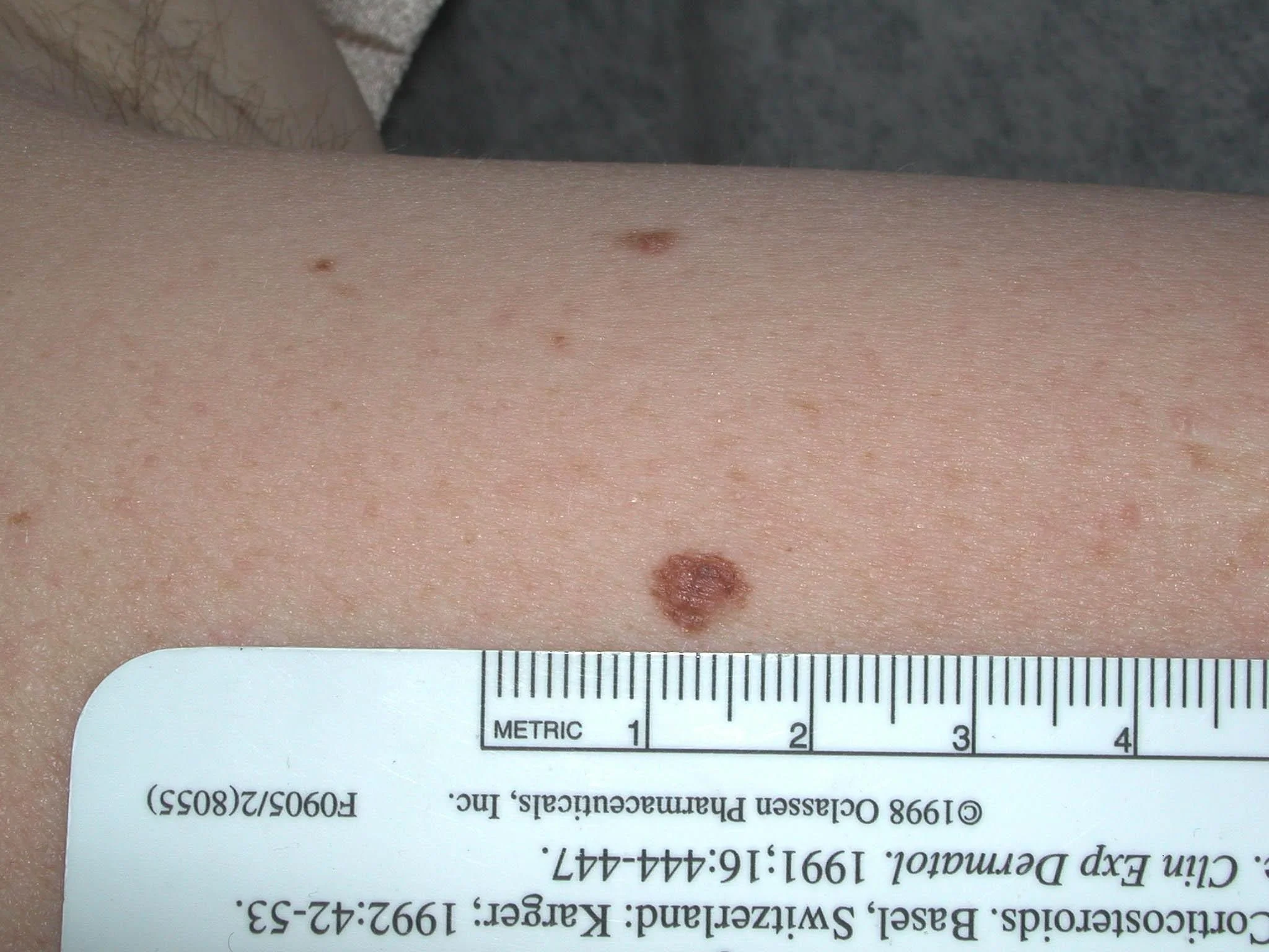
1-Ruler
- Accurately records the size of a lesion on successive examinations
- Measure in the longest axis first, then in the perpendicular axis
- e.g., this papule is 6x4 mm
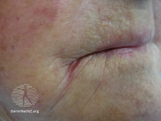
2- A penlight is used for side lighting
- Detects atrophy and fine
- wrinkling
- Distinguishes
- Flat from raised lesions
- Whether lesions are solid or fluid-
- filled
- Also helps look inside the mouth