The diagnosis of carcinoma of the cervix is normally made by cytology or biopsy and physical examination.
Endometrial carcinoma
may be suspected on ultrasound when there is widening of the endometrial stripe, but confirmation of the diagnosis is based on histology.
Magnetic resonance imaging
is useful to determine the extent of carcinoma of the cervix preoperatively.
- MRI is very accurate in assessing the local extent of the tumour.
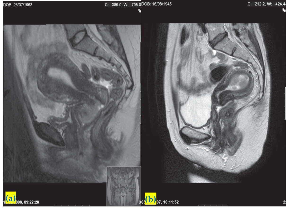
 (a)Sagittal T2-weighted MRI scan
showing a tumour confined to the cervix.
(b) Sagittal T2-weighted MRI demonstrating a polypoid tumour mass distending the endometrial cavity
CT and MRI
also enable the detection of dilatation of the ureters in cases where the tumour has caused ureteric obstruction.
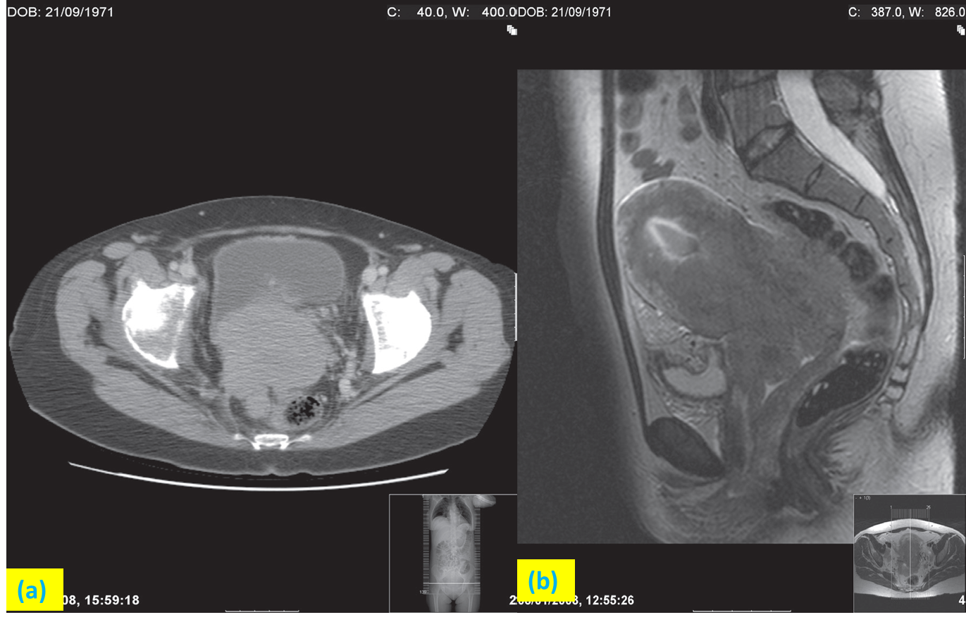
Advanced carcinoma of the cervix.
 (a)CT scan
showing a large tumour of the cervix extending into the rectum posteriorly.
(b) Sagittal T2-weighted MRI of the same patient. Note the tumour extending down the vagina.
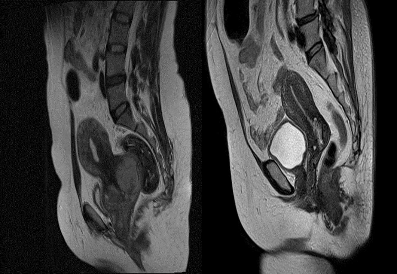
 Two different cases. T2 weighted sagittal MRI showing carcinoma of cervix