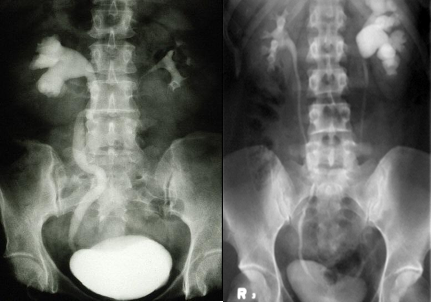
Distension and dilation of the renal pelvis and calyces.
It is usually caused by obstruction to the free flow of urine from the kidney.
If obstruction is at lower level, there is dilation of ureter and pelvis of kidney.
Untreated , initially it cause enlargement of kidney, but finally it leads to atrophy.

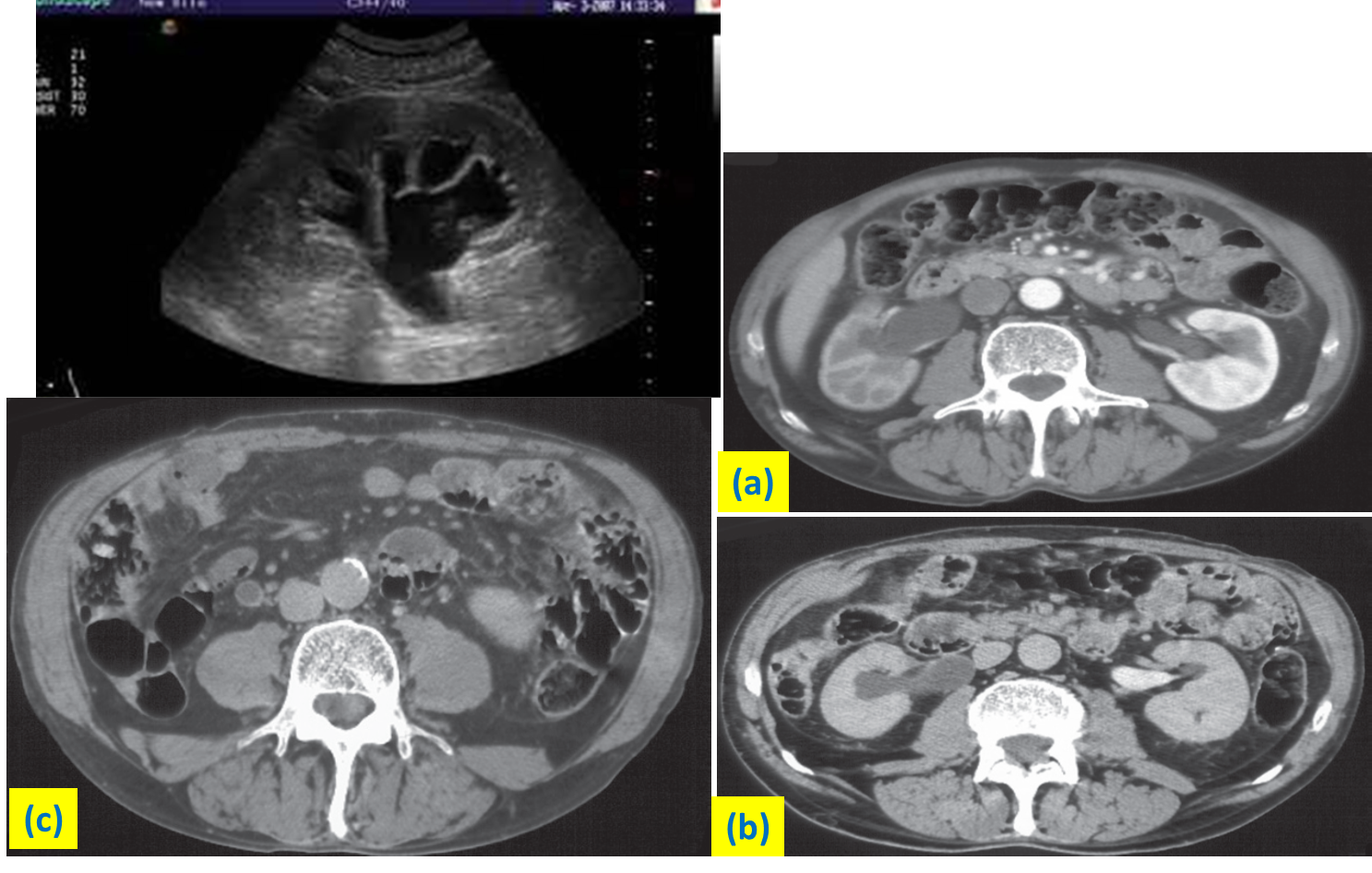
Ultrasound and CT images of hydronephrosis
(a) CT at the corticomedullary phase of enhancement. There is obstruction of the right kidney with dilatation of the pelvicaliceal system (b) CT at the delayed phase of enhancement. Intravenous contrast is seen in the left renal pelvis but not in the obstructed right renal pelvis. (c) CT through the dilated right ureter , in the same patient as (a) and (b).
