These are the commonest benign tumors during the reproductive age. They are often asymptomatic, but may cause menorrhagia ,infertility spontaneous abortion and postpartum hemorrhage. or as a palpable mass.
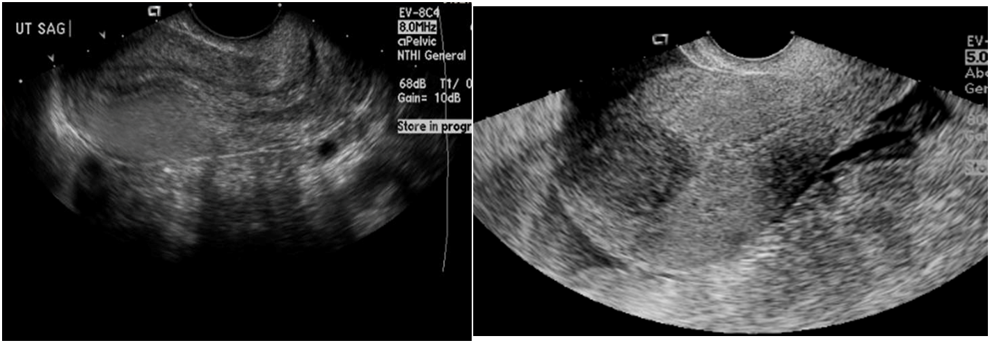
Ultrasound pelvis (transvaginal, transabdominal)
Most appropriate initial test for all patients with a suspected uterine leiomyoma
Supportive findings
- Well-circumscribed hypoechoic solid mass
- Calcifications and/or cystic areas due to degeneration
- Mass effect (e.g., hydronephrosis) may be seen in patients with large leiomyomas.
 Hypoechoic lesion in uterus is a typical appearance of a uterine leiomyoma
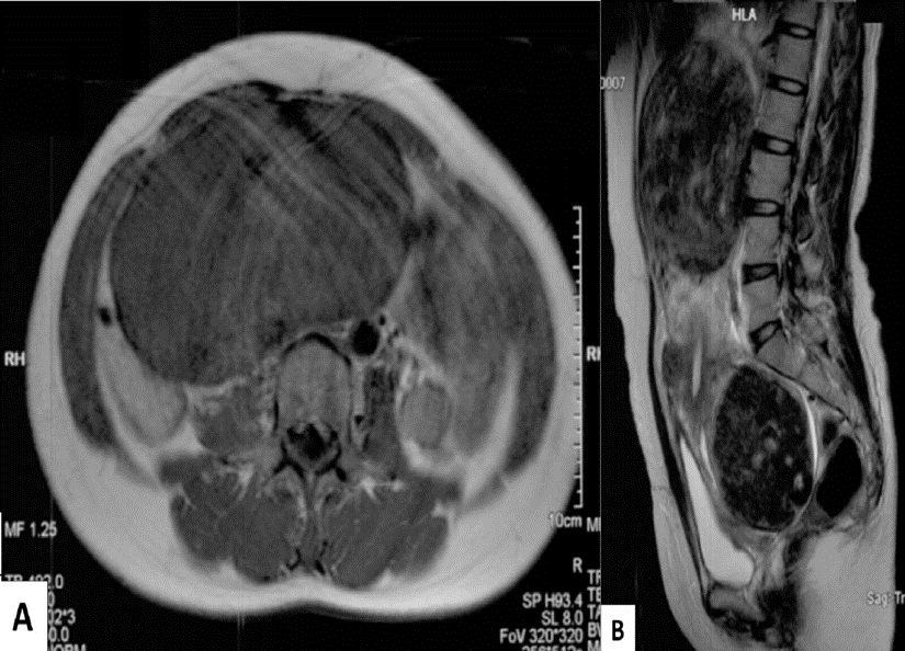
MRI pelvis without and with IV contrast
- Helps further characterize leiomyomas (e.g., before interventional procedures or surgery)
- Can rule out differential diagnoses of uterine leiomyomas
MRI abdomen and pelvis (A: T1 weighted, axial plane; B: T2 weighted, sagittal plane) of a 41-year-old female patient with a visible and palpable lower abdominal mass, hypermenorrhea, and pressure symptoms.
-
A: A large, smoothly circumscribed mass with moderate signal intensity can be seen.
-
B: The uterus is enlarged, with large heterogeneous masses in the area of the cervix and uterine fundus compressing the bladder and bowel loops.

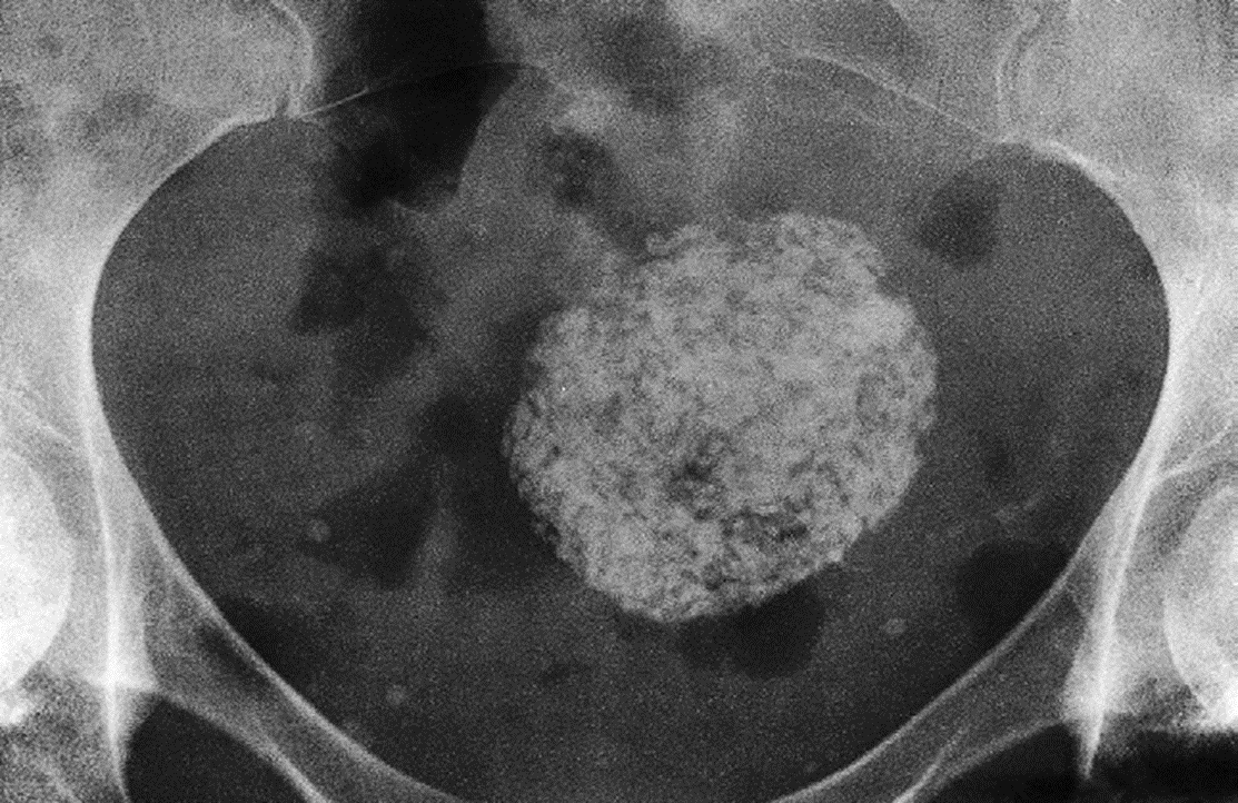
Calcified Uterine Leiomyoma Plain radiograph :
When sufficiently large, a fibroid can be seen on a plain film as a mass in the pelvis and may show multiple irregular but well-defined calcifications
Calcification in a large uterine fibroid.
