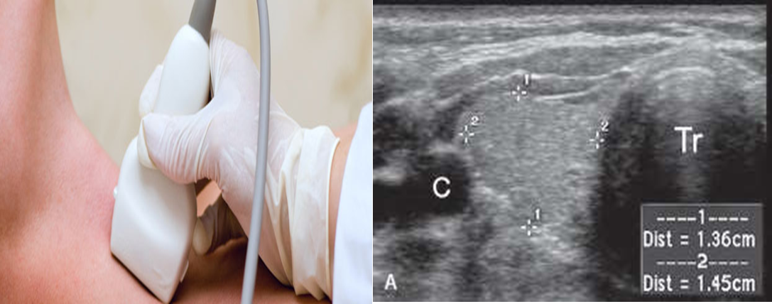
The thyroid is normally imaged by ultrasound.
Normal thyroid
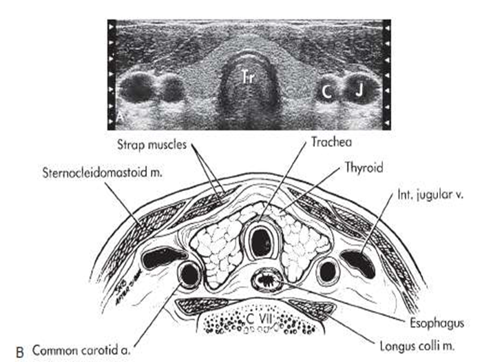
Ultrasound thyroid (transverse plane)
Normal image of the thyroid gland at the level of the isthmus.
1: right common carotid artery
2: right lobe
3: trachea
4: left lobe
5: left common carotid artery
6: left jugular vein
7: isthmus


Investigation of choice
-
Both right ,left lobes and isthmus are examined .Color Doppler is used to see vascularity. Ultrasound shows anatomical detail of thyroid and adjacent blood vessels and structures.
-
Normal parenchyma: homogeneous medium to high level echoes compared to adjacent neck muscles.
-
Ultrasound determines whether a nodule is cystic or solid or a mixture of both.
-
Simple cysts are invariably benign. Complex solid/ cystic lesions are usually benign. A solid mass could be a carcinoma or an adenoma.
