Endoscopic retrograde cholangiopancreatography (ERCP):
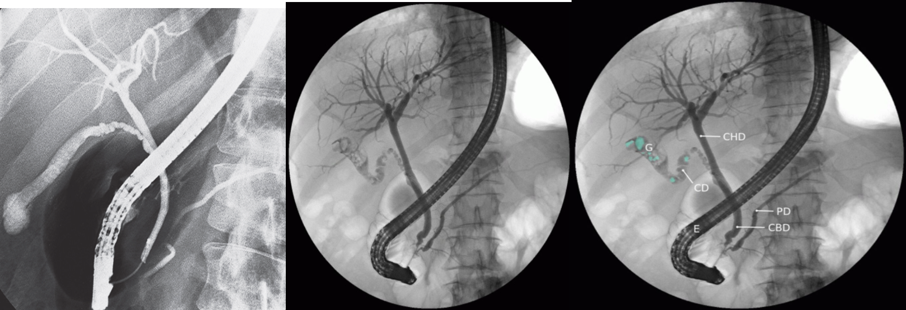
 ERCP showing cholelithiasis (first image is normal)
Endoscopic retrograde cholangiopancreatography (ERCP) The tip of the endoscope (E) is located within the duodenum at the ampulla of Vater. Contrast enhancement allows visualization of the hepatic biliary tract, left and right hepatic ducts, cystic duct (CD), gall bladder (G), common bile duct (CBD), and pancreatic duct (PD), CHD: common hepatic duct. Multiple filling defects (examples indicated by green overlay) can be seen within the gall bladder and cystic duct.
This finding is diagnostic of cholelithiasis.
Magnetic resonance cholangiopancreatography (MRCP):
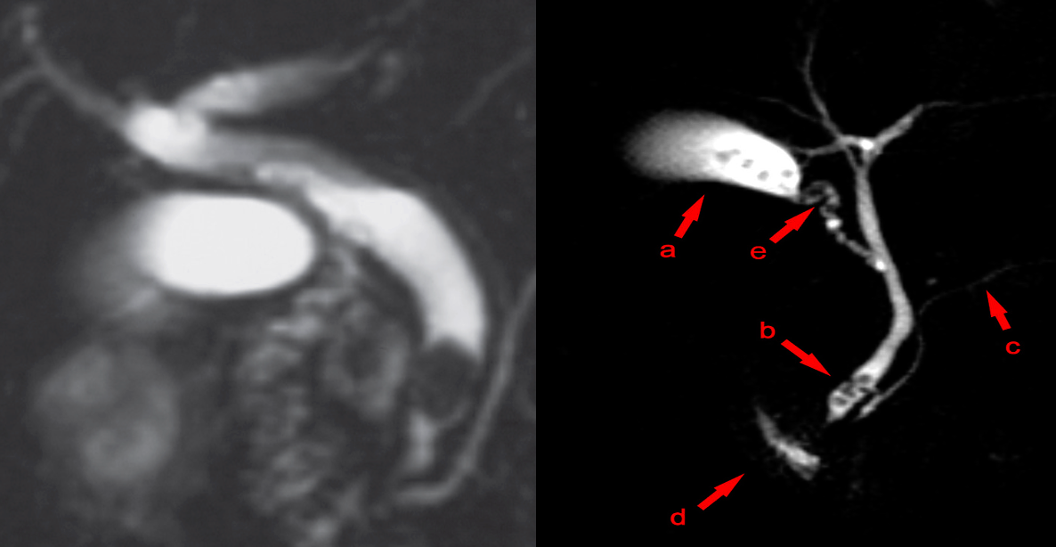
 MRCP showing cholelithiasis and Choledocholithiasis ((1st image normal))
Filling defects (i.e., the dark spots or areas that have not taken up the contrast) are visible within the gallbladder (a), the cystic duct (e), and the common bile duct (b). c: pancreatic duct; d: duodenum
These features are diagnostic of cholelithiasis complicated by choledocholithiasis.
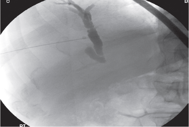
Percutaneous transhepatic cholangiogram (PTC):
