PET Scanner: positron emission tomography #z Mainly for following disease course, development, remission, etc…
- A PET scan uses a small amount of radioactive material (tracer). The tracer is given through a vein (IV). The tracer travels through the blood and collects in organs and tissues.
- The most commonly used agent is F-18 fluorodeoxyglucose (FDG).
- PET using FDG Z is the most sensitive technique for z staging solid tumours, such as bronchial carcinoma , and in the follow-up of malignancies, particularly lymphoma.
- PET is also used in the evaluation of ischaemic heart disease and in brain disorders such as dementia, epilepsy and Parkinson’s disease.
- A PET scan measure the important body functions such as blood perfusion of tissues, oxygen use and glucose metabolism.
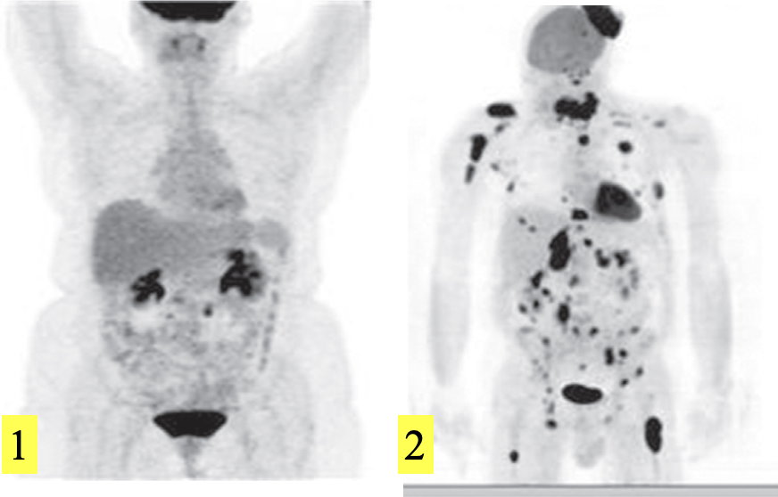
FDG-PET scans, maximum intensity projections.
-
Normal isotope distribution. There is intense uptake in the brain and the neck uptake is in the tonsils. The FDG is excreted by the kidneys.
-
Lymphoma, showing multiple visceral, nodal, bone and scalp deposits.

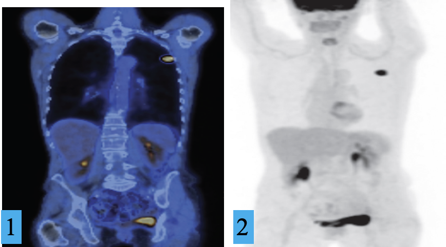
FDG-PET/CT of lung cancer. Z
1) Coronal CT image and
2) maximum intensity projection, demonstrating a small left lung cancer.
The remainder of the FDG uptake is physiological
