IM
Dr. Nada Abdelrahman
ITP

Overview
- Autoimmune disorder characterized by low platelet count (<100,000/mm3).
- Formerly known as Idiopathic Thrombocytopenic Purpura.
- Can be primary (isolated) or secondary to other conditions.
Immune thrombocytopenia: idiopathic thrombocytop
enic purpura (outdated): autoimmune disorder
Primary ITP: isolated thrombocytopenia (< 100,000/mm3) with no known precipitating
Secondary ITP: isolated thrombocytopenia secondary to an identifiable trigger such as SLE, APS, HCV, HIV.
Malignancy: lymphoma, leukemia (CLL)
Drugs: mn quinine, carbamazepine, heparin, vaccines, linezolid, sulfonamides, vancomycin.
Classification
- Newly diagnosed ITP: First 3 months.
- Persistent ITP: 3-12 months.
- Chronic ITP: > 12 months.
Pathophysiology
- Antiplatelet antibodies (mostly IgG directed against, e.g., GpIIb/IIIa, GpIb/IX) bind to surface proteins on platelets → sequestration by spleen and liver → ↓ platelet count → bone marrow megakaryocytes and platelet production increase in response (in most cases)
ITP is one of the most common autoimmune disorders. Platelets with antibodies on their surface are trapped in the spleen, where they are efficiently removed by splenic macrophages
Presentation
- Abrupt onset (childhood ITP)
- Gradual onset (adult ITP) common
- Asymptomatic (25% of cases): Found incidentally on routine blood tests. - Low platelet An incidental finding on a routine CBC in 25% of cases
- Symptomatic:
- Minor mucocut: Petechiae, purpura, Bruising tendency
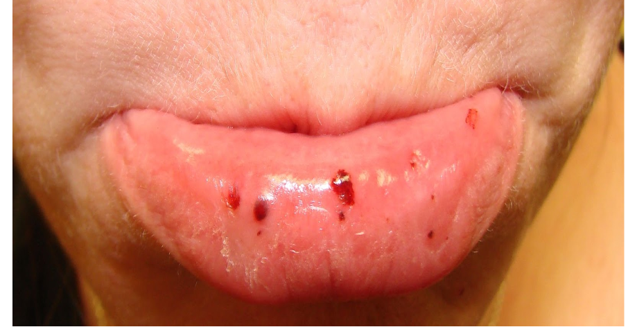
- Minor bleeding: Menorrhagia, epistaxis, gingival bleeding
- Catastrophic bleeding (rare): GI bleeding, intracranial hemorrhage.
Signs
- Spontaneous bleeding: platelet count < 20,000/mm 3.
- Retinal hemorrhages
- Purpuric rash
- Evidence of ICH, with neurologic symptoms
- Prolonged or excessive traumatic or surgical bleeding
- Splenomegaly is very unusual in ITP and makes other diagnoses more likely!
y Finally, most surgical procedures can be performed as long as the platelet count is above 50,000..
Platelets with antibodies on their surface are trapped in the spleen, where they are efficiently removed by splenic macrophages.
Diagnostic Considerations
Bleeding time is rarely measured nowadays, replaced by another screening tool called platelet function analyzers.
ITP is a diagnosis of exclusion; patients typically have a low platelet count with no other abnormalities. Routine
-
CBC: ↓ platelet count (< 100,000/mm3)
-
Coagulation panel: normal
-
Bleeding time: may be prolonged
-
Peripheral blood smear: N to large platelets
-
All adults: HIV and HCV screening
-
Bone marrow biopsy: atypical cases Findings: normal or ↑ megakaryocytes
-
Secondary ITP: antinuclear antibodies in SLE or H. pylori testing if the patient has GI symptoms or is from a high prevalence area
Diagnosis of exclusion.
- Lab Tests:
- Low platelet count
- Normal coagulation panel
- Peripheral blood smear: Normal to large platelets
- Bone marrow biopsy (in atypical cases): Normal or increased megakaryocytes
- Rule out secondary causes: HIV, HCV, SLE, H. pylori, medications.
ITP: Diagnostic Considerations Differential diagnosis
- Liver disease
- Lymphoproliferative
- Pregnancy-associated thrombocytopenia
- Infection/sepsis
- Acute leukemia
- Myelodysplastic syndrome
- Malignancy
- Megaloblastic anemia
- Transfusion
Treatment & Management
All pt; stop medications (NSAIDS), and ttt underline
-
Observation: only asymptomatic adults with a platelet count >=30 * 109/L.
-
Minor symptoms : oral prednisolone as first choice.corticosteroids
-
Second choice (contraindication, non-response:) IVIG or anti-Rho(D) immunoglobulin (risk of intravascular hemolysis)
-
Life-threatening bleeding: critical care.
-
Refractory cases: high-dose parenteral glucocorticoids and IV immunoglobulin (IVIg), with or without platelet transfusions.
-
IV immunoglobulins: increase platelet survival
-
Platelet transfusion is indicated for controlling severe hemorrhage prior to surgery.
Newly-diagnosed ITP refractory to first-line medical therapy, or persistent/chronic ITP.
-
Thrombopoietin receptor agonists (TPO-RAs): increase platelet production by stimulating megakaryocytes in the bone marrow : Romiplostim,
-
Rituximab monoclonal antibody targeting CD20 - immunity
-
Splenectomy Indications
- Treatment-resistant thrombocytopenia lasting > 12 months
*Emergency splenectomy: *
- with life-threatening bleeding in whom medical therapy fails.
Case: Suspected ITP
A 42-year-old female presented with gingival bleeding and dark purple spots on her legs. The patient first noticed the dark spots on her legs 2 months ago, but the gingival bleeding started 5 days ago. Past history is notable for intravenous drug use and hepatitis C infection. Normal vital signs. Examination of the patient’s oropharynx and gingiva reveals petechiae with pinpoint bleeding. Cardiac, pulmonary, and abdominal exams are noncontributory. Purpura are observed on the bilateral legs. Laboratory testing is obtained, and the results are shown below.
- Platelet count 25,000/mm3
- Hemoglobin (Hb) 13.5 g/dL
- Leukocyte count 11,000/mm3
Which of the following serological findings would most likely be present in this patient?
Answer: CC
Surgery
Idiopathic Thrombocytopenic Purpura
ITP
-
ITP in children:
- 2-4 years age
- Acute
- Usually post viral,
- Most recover without treatment.
-
ITP in adults:
- Chronic
- Antibody (IgG) against platelets
- Low platelets <50,000 (epistaxis, GI bleeding, ecchymosis)
- Mild splenomegaly.
- Initial therapy (if bleeding)- prednisolone, platelet concentrate, & immunoglobulin.
- Splenectomy: (Commonest elective indication)
- Persistent < 30,000 platelet after 4-6 weeks of medical therapy.
- Severe thrombocytopaenia- platelet concentrate given after splenic artery ligation. Long time remission in 65%
- 2nd line therapy- Rituximab (anti-CD20 monoclonal antibody)