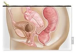
Female Genital Tract & Pelvic Floor
By Dr. Mona Ahmed
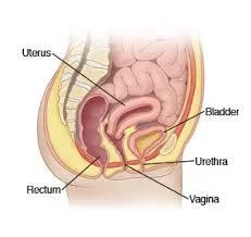
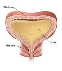
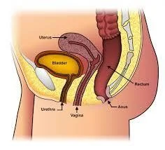
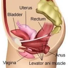
The Bladder
- The bladder wall is made of involuntary muscle.
- It is lined with transitional epithelium.
- Has an average capacity of 400 ml.
- The ureters open into the base of the bladder.
- The urethra leaves the bladder below the ureteric orifices.
- The base of the bladder is adjacent to the cervix & separated from the anterior vaginal wall by the pubocervical fascia.
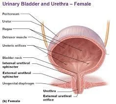
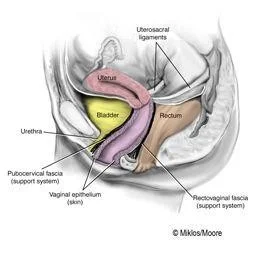
The Urethra
- The female urethra is 3.5 cm long.
- and lined with transitional epithelium.
- The lower part occasionally become infected.
- The upper part of the urethra is mobile but the lower part is fixed.
The Ureter
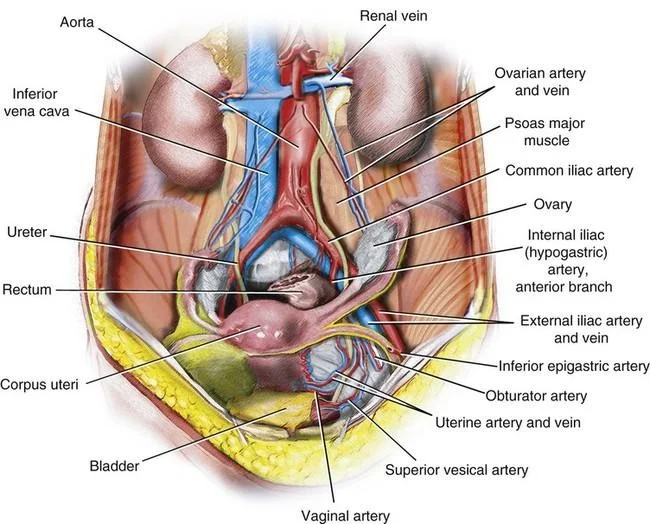
- As the ureter crosses the pelvic brim
- it lies in front of the bifurcation of
- the common iliac artery.
- The ureter runs close to the lateral vaginal fornix to enter the bladder.
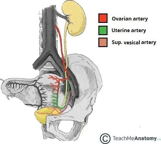
- Blood supply from the ovarian artery.
- Due to its close relationship to the cervix
- the vagina and the uterine artery
- the ureter may be damaged during hysterectomy (cut or tied).
The Rectum
- The rectum extends from the level of the third sacral vertebra to the anal canal.
- The front and sides are covered by the peritoneum of the rectovaginal pouch.
- The middle third only the front is covered by peritoneum.
- Rectum is separated from the posterior wall of the vagina by the rectovaginal fascial.
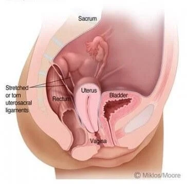
- Lateral to the rectum, the uterosacral ligaments.
The Pelvic Muscles, Ligaments and Fascia
The Pelvic Diaphragm
- The pelvic diaphragm is formed by the levator ani muscles, which are broad, flat muscles the fibres.
- The two muscles, one on either side, constitute the pelvic diaphragm.
- The muscles arise from:
- The body of the pubis.
- The pelvic fascia.
- The ischial spine.
- The levator ani muscle inserted into:
- The preanal raphe.
- The anal canal.
- The anococcygeal raphe.
- The coccyx.
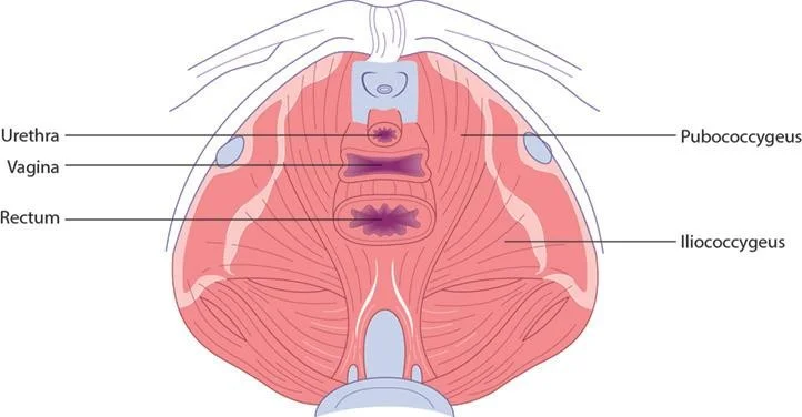
Parts of Levator Ani Muscle
The muscle is described in two parts:
- The pubococcygeus.
- The iliococcygeus.
They have some sphincteric action. The nerve supply is from the third and fourth sacral nerves. The pubococcygeus muscles support: - the pelvic - abdominal viscera (including the bladder). Together with the fibres from the opposite muscle they form a loop that maintains the angle between the posterior aspect of the urethra and the bladder base. During micturition, this loop relaxes to allow the bladder neck and upper urethra to open and descend.
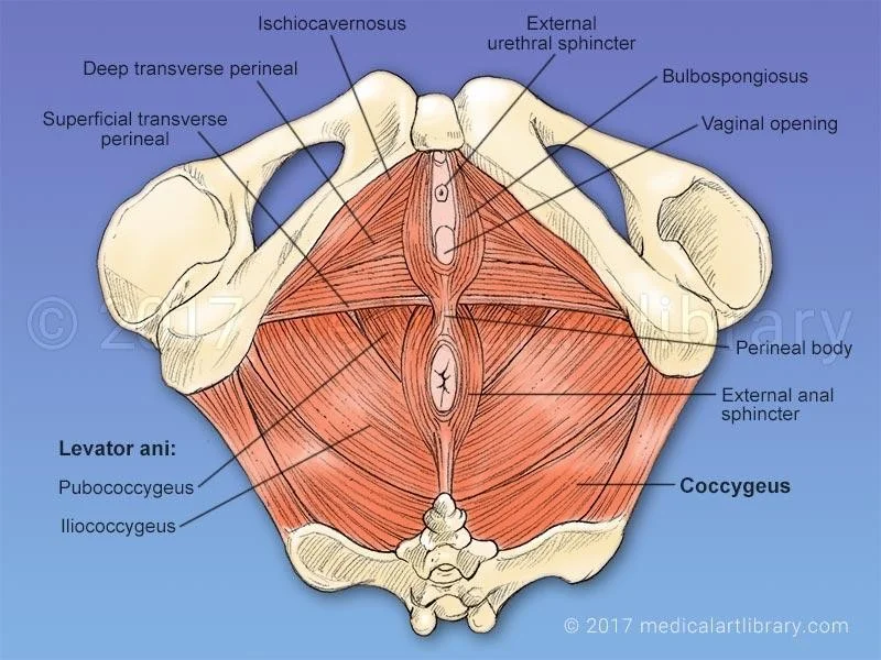
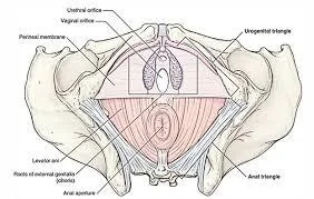
Urogenital Diaphragm
- Is made up of two layers of pelvic fascia.
- The deep transverse perineal muscles (compressor urethrae) lie between the two layers and the diaphragm is pierced by the urethra and vagina.
- The perineal body This is (a mass of muscular tissue) that lies between the anal canal and the lower third of the vagina.
- Its apex is where the rectum and posterior vaginal walls come into contact.
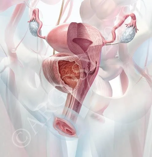
The Pelvic Peritoneum
The Broad Ligament
- peritoneum double reflection from the lateral borders of the uterus on either sides.
- Its not a ligament but a peritoneal fold and it does not support the uterus.
- The Fallopian tube runs in the upper free edge of the broad ligament.
- The lateral portion is called the ‘infundibulopelvic fold’ “suspensory” where ovarian vessels and lymphatics.
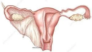
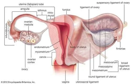
The Mesosalpinx
- the portion of the broad ligament that lies above the ovary.
The Mesovarium
- is a short mesentery that attached the ovary to the posterior layer of the broad ligament through which the ovarian vessels lies.
The Ovarian Ligament
- passes from the medial portion of the ovary to the uterus.
The Round Ligament
- continuation enter the inguinal canal, to end in the the labium major.

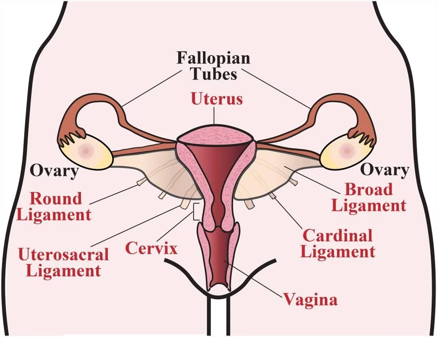
Uterine Support
- ➤ The cardinal ligaments (transverse cervical ligaments)
- ➤ The uterosacral ligaments run from the cervix and vagina to the sacrum.
- ➤ Round ligament
- The bladder is supported by:
- ✓ pelvic fascia.
- ✓ pubocervical fascia.
The Blood Supply
The Ovarian Arteries
- supply the ovary and tube
- they anastomose with the terminal
- branches of the uterine artery.
The Internal Iliac (Hypogastric) Artery Branches
- supply the pelvic organs by the following branches:
- The uterine artery …supply to the uterus.
- The vaginal artery …supply the vagina.
- The vesical arteries … supply the bladder and terminal ureter.
- The pudendal artery… supplying the vestibular bulbs and clitoris.
- The superior rectal artery …supply the rectum.
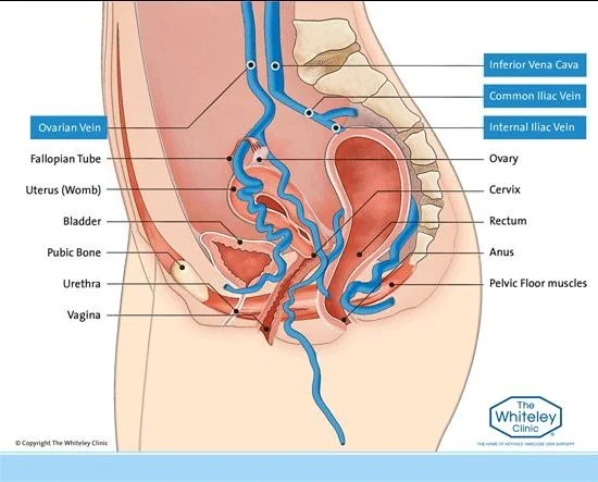
The Pelvic Veins
- ✓ Around the bladder
- ✓ Uterus
- ✓ vagina
- ✓ rectum form… plexuses that drain into the internal iliac veins.
- ✓ The ovarian veins on each side begin in the pampiniform plexus in the broad ligament ending in the inferior vena cava.
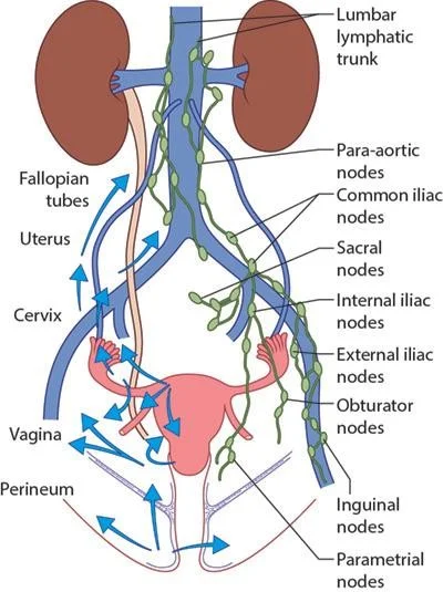
Lymphatics
- The pelvic lymphatics draining:
- inguinal nodes.
- superficial femoral nodes.
- Pelvic nodes.
- para aortic nodes.
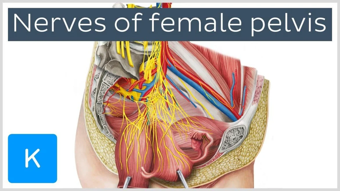
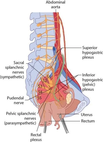
The Nerves
Nerve Supply (Somatic)
- The pudendal nerve.
- The perineal nerve.
- sacral nerves.
Autonomic Nerve Supply
- of the pelvic viscera:
- the preaortic plexus
- superior hypogastric plexus