Burns & Scalds
Introduction
Thermal injuries are caused either due to exposure to cold or exposure to heat. The effect of heat or cold over the body may be in generalized form or may have localized effects on a particular body part. Thus, thermal injuries are classified as follows:
A. Due to Exposure to Cold
- Hypothermia
- Frostbite
- Trench foot
- Chilblains
B. Due to Exposure to Heat
- Heat stroke
- Heat cramps
- Heat exhaustion
- Burns
- Scalds
Burns
Definition
Burn is defined as local injury caused by the application of heat or chemical substance to the external or internal surface of the body, which causes destruction of tissue. The minimum temperature for producing a burn is about 44 ºC for long enough to damage the tissue.
Pathophysiology
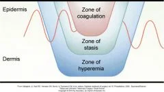
Burn injury causes coagulative necrosis of the epidermis and underlying tissues. Burns bring cellular damage primarily by transfer of energy inducing coagulative necrosis. Chemicals and electricity cause direct injury to cell membrane in addition to the transfer of heat. The burn injury of the skin has been divided into three zones of coagulative necrosis
Degrees of Burn
A. Dupytren’s Classification
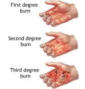
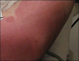
- First degree – it is a superficial burn involving only epidermis with a low degree of heat for a small or short period.
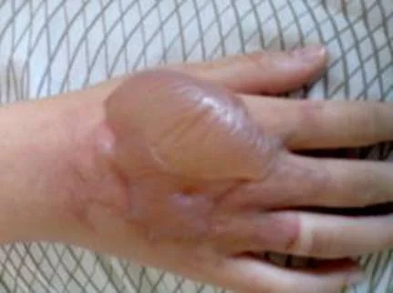
- Second degree – burns are limited to the epidermis. There may be blister formation.
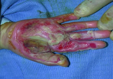
- Third degree – epidermis is destroyed completely.
- Fourth degree – burns involve dermis.
- Fifth degree – burn depth extends to subcutaneous (hypodermis) tissue. The lesion is less painful due to destruction of nerves.
- Sixth degree – burn lesion extends deeper to involve muscles and bones.
B. Wilson’s Classification
- First degree — epidermal burns, it includes first and second degree of Dupytren’s classification. Blisters may form and the lesion may be painful.
- Second degree — dermoepidermal burns, it includes third and fourth degree of Dupuytren’s classification. The lesion is painful and characterized by blister formation.
- Third degree — deep burns, it includes fifth and sixth degree of Dupuytren’s classification. The burns are less painful due to nerve destruction.
C. Modern Classification Y
- Superficial burns - it is dermo-epidermal burn injuries and here burns do not extend the full thickness of the skin.
- Deep burns - here the burn lesion involves deeper structures beneath the skin. Any burns involving more than true skin are grouped in this category.
Estimation of Extent of Burn
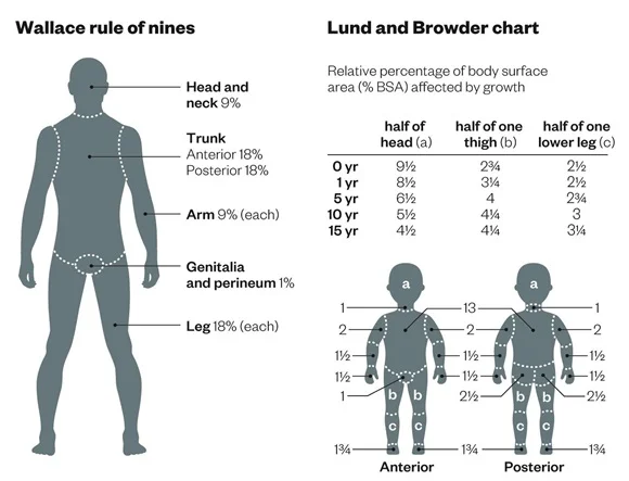
Wallace Rule of Nines
- Head and neck 9%
- Trunk
- Anterior 18%
- Posterior 18%
- Arm 9% (each)
- Genitalia and perineum 1%
- Leg 18% (each)
= 100%
Lund and Browder Chart
Relative percentage of body surface area (% BSA) affected by growth.
| Age | half of head (a) | half of one thigh (b) | half of one lower leg (c) |
|---|---|---|---|
| 0 yr | 9½ | 2¾ | 2½ |
| 1 yr | 8½ | 3¼ | 2½ |
| 15 yr | 6½ | 4 | 2¾ |
| 10 yr | 5½ | 4¼ | 3 |
| 15 yr | 4½ | 4¼ | 3¼ |
Cause of Death
Immediate
- Neurogenic shock
- Hypovolemic shock
- Inhalation of smoke & CO
- Laryngeal edema of glottis
Delayed
- Septic shock - the only Shock
- Toxemia
- Renal failure
- Pulmonary embolism (rare)
- Fat embolism (rare)
- ARDS
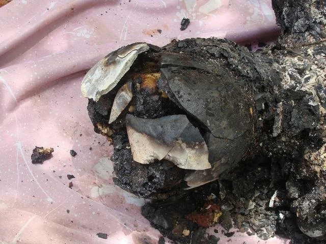
Autopsy Findings
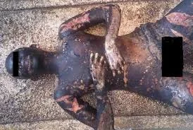
1. External Examination
- Heat stiffening or heat rigor
- Boxer’s attitude or pugilistic attitude
- Contraction of the paraspinal muscles often causes a marked opisthotonos
- Hairs are either singed or completely burnt away
- In severe burns or sustained application of heat, the muscle or even bone may be charred
- If death occurs some days after the incident, there will be presence of pus and necrosis in the burn areas. There may be ulceration and sloughing of tissue
2. Internal Findings
- Blood may be bright if death is associated with carbon monoxide inhalation
- Internal organs are congested or may show partly cooked or cooked appearance
- Soot particles or carbon particles may be seen in the respiratory passage due to smoke inhalation
- Bones may show heat fracture or thermal fracture
- Heat hematoma. This occurs in the head if the victim has been subjected to intense heat for a longer duration
Difference Between Antemortem and Postmortem Burns
| Features | Antemortem Burns | Postmortem Burns |
|---|---|---|
| Line of redness | Present around burn area | Absent |
| Blisters | Present and contains serous fluid rich in protein and chlorides. The base is red, inflamed with raised papilla | Usually absent and if present contains air and clear fluid. The base is dry, pale or yellow, hard and horny |
| Vital reaction | Present | Absent |
| Reparative process | Present | Absent |
| Infection | Present | Absent |
| Carbon/soot particles in respiratory passage | Present | Absent |
| Carboxyhemoglobin in blood | Present | Absent or low level |
| Cyanide in blood | Present | Absent or low level |
Medicolegal Importance in Burn
- Questioning about the identity of the deceased
- Whether the burns are antemortem or postmortem? And what was the time of sustaining burns injuries?
- Was it the cause of death? Are the antemortem burns sufficient to cause death?
- And most importantly, were the burns suicidal, homicidal, or accidental? Bride burns and dowry death
Scald
Definition
Scalds are produced by moist heat or any hot liquid, such as:
- Water
- Oil
- Or even molten metal
Characteristics
- Less severe than burns produced by dry heat unless the liquid is superheated
- It appears: erythematous with desquamation and blistering
- Generally occur on exposed skin
- The pattern of the scald can give an indication of how it occurred
Statistically, Scalding is the Most Common Type of Thermal Injury Among Children
- Can cause hypovolemic shock, electrolyte disturbances, and secondary infection which may lead to death
- Inhalation of moist steam may also lead to death because of Asphyxia
Types of Scalds
Liquid Scalds
- Hot water, including tap water in bathtubs and showers, is the leading cause of both scalds and hospital admissions for burns
Intentional Scalding
-
Intentionally inflicted scalding can be a component of child or elder abuse
-
The pattern of the injuries can be very important in determining that the burns are inflicted rather than inadvertent
Degrees of Burns
The image illustrates three degrees of burns, each with distinct characteristics:
- 1st Degree: Erythema formation above the affected area
- 2nd Degree: Blister formation with increased vascular permeability
- 3rd Degree: Drying and desiccation of the underlying tissue with necrosis
Features of Scald
- Have sharply demarcating edge, correlative to the harmful agent
- They also don’t cause any charring or singeing of surface hairs
- Trickle pattern comes along as a sign of splashing of hot fluid causing the effect of gravity
- A horizontal level may be made by the hot fluid if hands are immersed in a bucket full of it
Differences Between Burns & Scalds
| Features | Burns | Scalds |
|---|---|---|
| Site | Injury occurs at/above the site of application of causative agent | Injury occurs at/below the site of application of causative agent |
| Cause | Dry heat | Moist heat |
| Skin | Reddening to superficial burns to charring | Erythema, blister, may be sodden & bleached |
| Scar | Thick & contracted | Less contracted |
| Charring of skin/Singeing of hairs | Present | Absent |
| Splashing | Absent | Present |
| Clothes | May show evidence of burn like melting of fibers | Not burnt but may be wet |
Medicolegal Importance of Scalds
-
Scalds are usually accidental, involving infants, toddlers, or children
-
They may be intentional, such as abusing, torture, and some with malicious intention may use it as a means of retaliation
-
It’s also may be a sign of battered baby syndrome; for those reasons, we must be alert to it
Chemical Burns
- Chemical burns are produced by corrosive acids and alkalis
- The pattern on the skin surface resembles that of scalds caused by liquids, but the injuries differ in physical appearance, reflecting different mechanisms of tissue damage
- The amount of tissue damage caused in a chemical burn will depend on the strength of the chemical
- Chemical burns from acids and alkalis are almost invariably accidental, and suicide by the ingestion of strong acid or alkali has now become rare

Electrical Burns
-
Skin burns are a common form of electrical injury
-
A firm contact electrical burn of entry typically leaves a central collapsed blister x
-
The severity of the electrical injury to the deep tissues depends on the amperage

Lightning
- Lightning bolt is produced when the charged undersurface of a thundercloud discharges its electricity to the ground
- Very large electrical forces are involved
- The lightning may directly strike the victim
- The resulting injuries may be electrical, burns, or blast from the wave of heated air created by the lightning strike
- An unmarked dead body found in the open should raise the possibility of a lightning strike


Thermal Injury Terminology
Cherry Red Lividity
- In individuals with a significant carboxyhemoglobin concentration in the blood (due to carbon monoxide exposure), the livor mortis has a distinctive, bright, cherry red color

Epidural Hematoma
- In burned bodies, epidural hematoma results from blood and marrow in the cancellous diploe of the skull boiling and being extruded by the intense heat
Fire Fractures
- Fire fractures are another postmortem heat-related artifact and should not be confused with antemortem injuries. Because of the extreme heat, the muscle, tendons, and soft tissue are charred
Lichtenberg Figure, Ferning, Keraunopathy
- Keraunopathy is most generally the study of lightning-related injury, and the term is also used occasionally as synonymous with ferning, the arborizing pattern of skin discoloration seen on some bodies after a lightning strike
Pugilistic Attitude
- The burned body assumes what appears to be a boxer’s stance. This is a postmortem artifact resulting from the effects of heat on the muscle protein

Skin Splits
- Skin splits are usually seen on or near joint surfaces, the extensor surfaces of the extremities, and the head. These splits can extend to the underlying adipose and skeletal muscle, dependent on the intensity of the heat

Soot
- Soot is an amorphous black material found in the airways (mouth, nares, trachea, bronchi) of individuals inhaling smoke during a fire

Histology and Microscopic Examination
-
Histologic examination of the edge of a burn, to include both the burned area and adjacent grossly unburned skin, may reveal vital reaction microscopically, consisting of acute inflammation, hemorrhage, edema, and necrosis
-
These findings are present only in antemortem burns


