Is a common incidental finding and rarely requires treatment.
Occasionally, they can cause significant haemorrhage, especially following trauma, and therefore percutaneous biopsy should be avoided if possible.
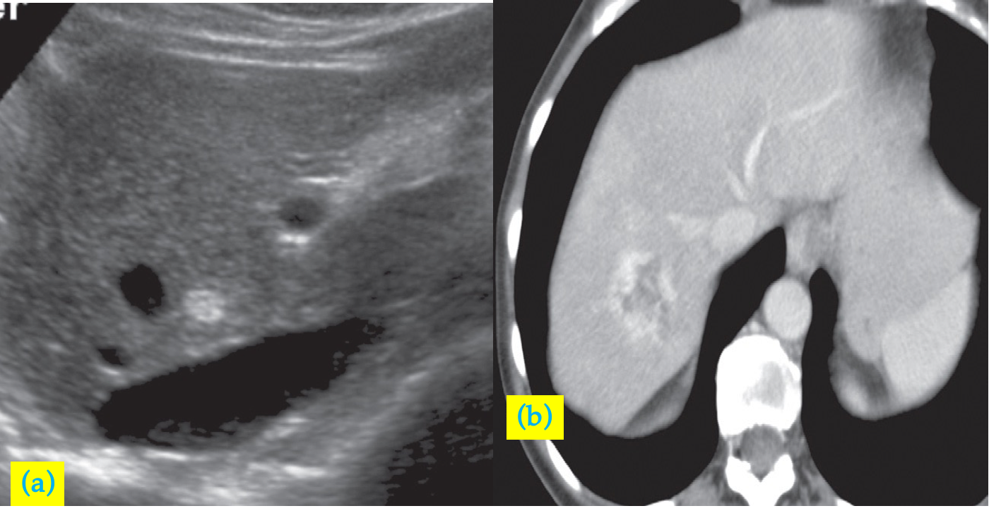
At ultrasound, haemangiomas are typically well-defined, peripheral, echogenic masses z
At CT, there is usually a characteristic enhancement pattern characterized by sequential contrast opacification, beginning as nodular or globular areas of enhancement at the periphery and proceeding toward the centre over time until the density increases to become similar to that of the surrounding liver.
 Haemangioma (incidental finding).
- (a) Ultrasound scan showing an echogenic mass in the right lobe of the liver . IVC, inferior vena cava.
- (b) CT scan, in another patient, after intravenous contrast enhancement showing a low density lesion in the right lobe of the liver with peripheral nodular enhancement, characteristic of a haemangioma.