Investigations
Non-Contrast Computed Tomography (CT) Brain
- Consider if the patient has:
- Sudden onset severe headache that reaches maximal severity within one hour.
- Headache with ≥1 of the following red flags:
- Increasing frequency or severity
- Fever or neurological deficit
- History of cancer or immunocompromise
- Older age (>50 years) of onset
- Post-traumatic onset
- Headache with new onset or pattern during pregnancy or peripartum period
- Headache with features of intracranial hypertension (e.g., papilledema, pulsatile tinnitus, visual symptoms worse on Valsalva).
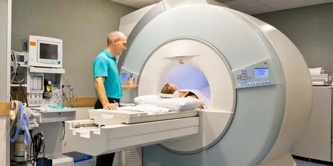
MRI Brain
- Consider if the patient:
- Headache with features of intracranial hypertension (e.g., papilloedema, pulsatile tinnitus, visual symptoms worse on Valsalva).
- Headache with features of intracranial hypotension (e.g., positional, worse when upright, better when lying down).
- Headache with new onset or pattern during pregnancy or peripartum period.
- Headache with one or more of the following red flags:
- Increasing frequency or severity
- Fever or neurological deficit
- History of cancer or immunocompromise
- Older age (>50 years) of onset, or post-traumatic onset.
- New primary headache of suspected trigeminal autonomic origin.
Lumbar Puncture (LP)
- Order an LP after a negative CT without contrast:
- If the patient has the worst headache of their life, or a ‘thunder-clap headache’ (SAH)
- If the patient has a fever (brain abscess, meningitis, encephalitis)
- If the patient has neck stiffness (SAH, meningitis)
- If the patient is young, overweight, and female (sinus venous thrombosis, pseudotumor cerebri)
- After negative CT when considering SAH, meningitis, pseudotumor cerebri.
Laboratory Tests
- Erythrocyte sedimentation rate, when considering giant cell arteritis
- ABG, when considering hypoxia or hypercapnia
- Carboxyhaemoglobin, when considering carbon monoxide poisoning
- A pulse CO-oximeter may reveal elevated CO levels, but this test is not widely available.
- FBC and liver function tests are performed if pre-eclampsia is suspected. Urinalysis is also required in these patients.
Limit of Imaging
Limit of Imaging
SNOOP
-
Systemic symptoms, illness, or condition (e.g., fever, weight loss, cancer, pregnancy, immunocompromised state including HIV)
-
Neurologic symptoms or abnormal signs (e.g., confusion, impaired alertness or consciousness, papilledema, focal neurologic symptoms or signs, meningismus, or seizures)
-
Onset is new (particularly for age >40 years) or sudden (e.g., “thunderclap”)
-
Other associated conditions or features (e.g. head trauma, illicit drug use, or toxic exposure; headache awakens from sleep, is worse with Valsalva maneuvers, or is precipitated by cough, exertion, or sexual activity)
-
Previous headache history with headache progression or change in attack frequency, severity, or clinical features.
Secondary Headache Source
-
Impaired vision or seeing halos around light (glaucoma)
-
Subacute angle closure glaucoma.
-
Visual field defects (lesion of the optic pathway e.g. pituitary mass).
-
Sudden, severe, unilateral vision loss (optic neuritis)
-
Morning headache is nonspecific (primary headache syndrome, sleep apnea, chronic obstructive pulmonary disease, obesity hypoventilation syndrome)
-
The presence of nausea, vomiting, worsening of headache with changes in body position (particularly bending over), an abnormal neurologic examination, and/or a significant change in prior headache pattern (tumor).
-
Intermittent headaches with high blood pressure (pheochromocytoma).
MRI vs. CT
-
Detecting edema
-
Vascular lesions
-
Intracranial pathology
(posterior fossa) -
Available
-
Urgent or Emergency care
-
Concern for subarachnoid hemorrhage
(thunderclap headache)