Viral Skin Infections CS-OSPE
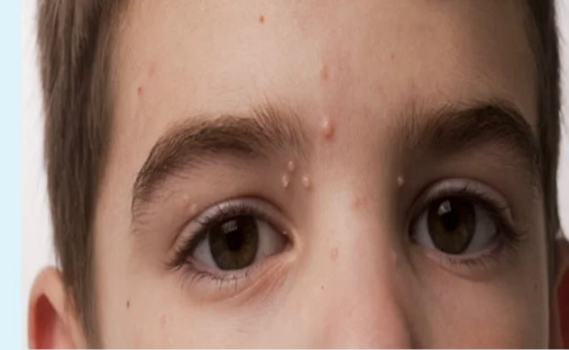
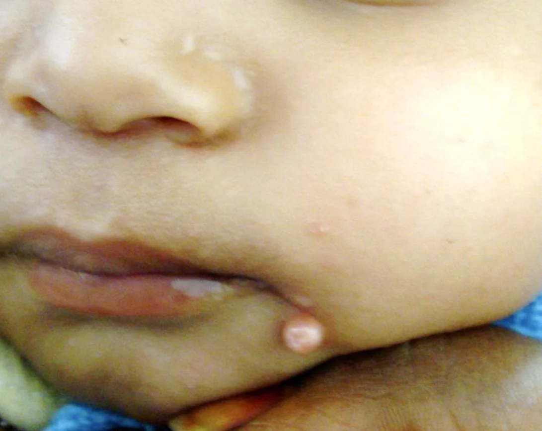
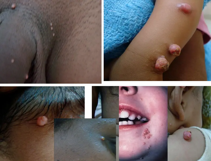
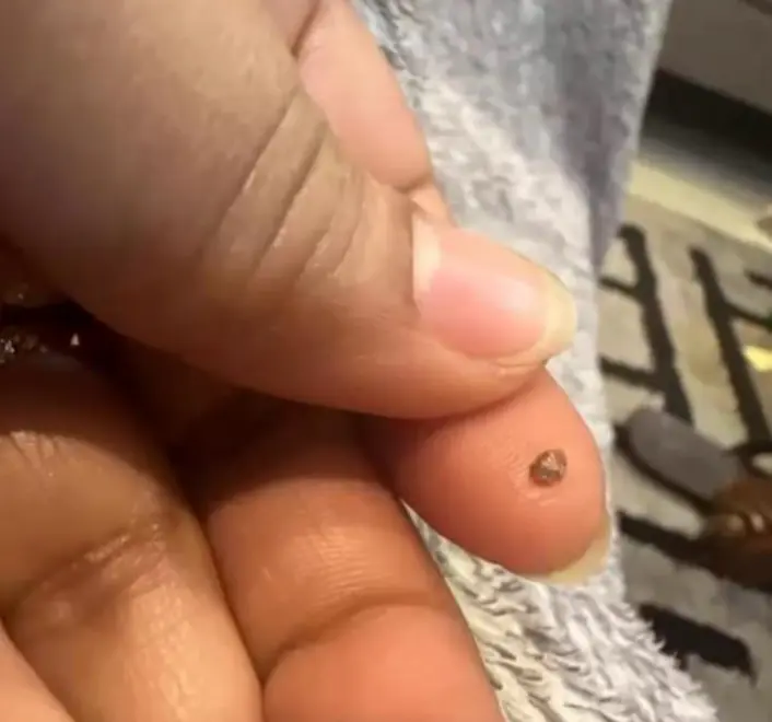
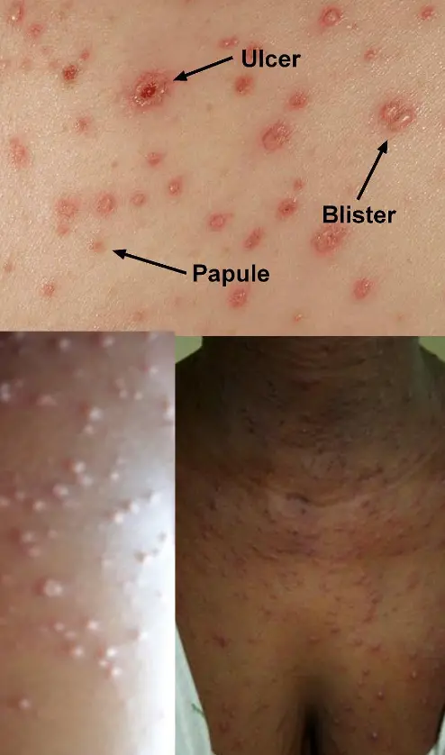
Molluscum Contagiosum
Diagnosis: Z
- Molluscum Contagiosum
DDx / Differential Diagnoses:
- Warts
- Chickenpox
Causative agent/organism:
- Poxvirus
Transmission:
- In this case, sexual
Clues that support diagnosis:
- Young age, painless, and not itchy.
- Started as single, small papules then increased in numbers and sizes.
- Infant developed this disease 2 weeks after she joined a day care.
Clinical Presentation
- Smooth.
- Shiny.
- Skin colored.
- Central depression.
- Multiple papule or nodules.
Question that helps to reach a diagnosis:
- Does anyone in the school have the same condition?
Treatment options/Best treatment:
- Child: self limiting within 1 year.
- Physiological destruction: Z
- Curettage: warts are anesthetized and then scraped with a curette.
- Cautery: electrical burning of lesions.
- Cryotherapy: by Liquid nitrogen.
- Surgical removal or excision.
- Laser: is used also for removal of warts.
- Chemical destruction:
-
- Chemical: Salicylic acid, Silver nitrate (25% for children, 75% for adults) Z
- Salicylic acid: in high concentrations.
- Silver nitrate: in form of pencils.
-
Most likely HIV+ patient
 ??
??
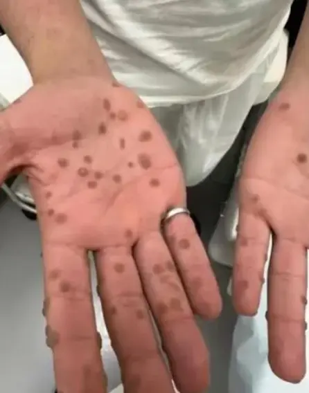
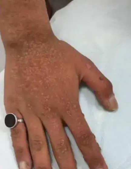
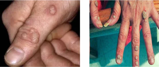
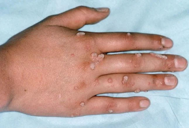
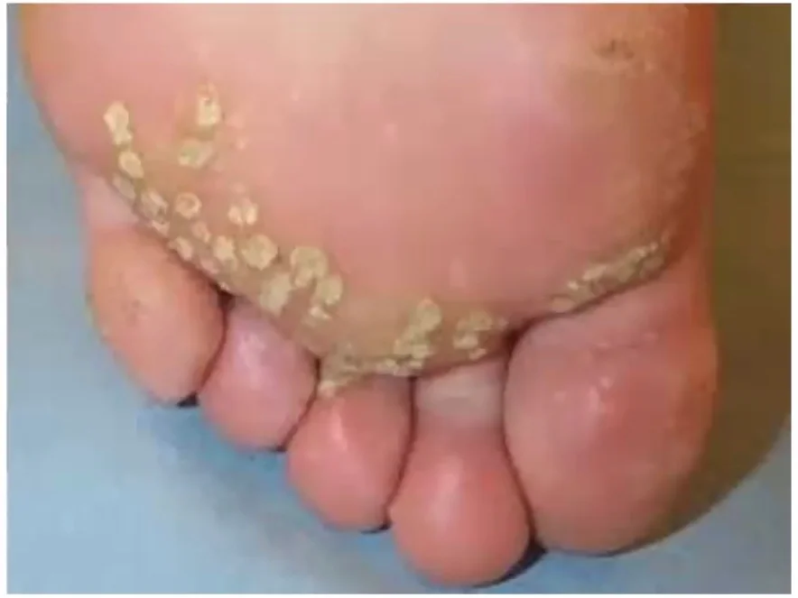
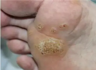
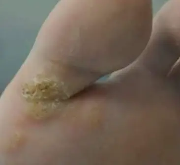
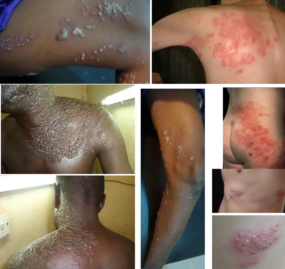
Warts (Verrucae): Common, Plantar, and Flat Warts
Diagnosis:
- Warts (Verrucae): Common viral wart (verruca vulgaris), Flat wart (verruca plana), Plantar wart (verruca plantaris)
Describe:
- Extensive because patients have immunodeficiency. Usually papular, but may appear macular.
Causative agent:
- Human papilloma virus (HPV) subtypes 36 - 38 Z severe forms 16 18
Types:
- Common wart = verruca vulgaris
- Flat wart = verruca plana
- Plantar wart = verruca plantaris
Differential diagnosis:
- Seborrheic keratoses
- Dermatosis papulosa nigra
- Nevi
- Pityriasis versicolor
- Psoriasis (plaque)
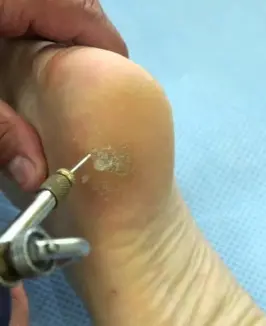
Management/Treatment:
- Full history
- Examination
- Education
- Tissue destructive modalities:
- Keratolytic (salicylic acid and podophyllin)
- Cryotherapy (Liquid nitrogen(-196C°))
- Electrotherapy (ED)
- CO2 laser
- Cautery
- Curettage
- Silver nitrate
- Immunotherapy
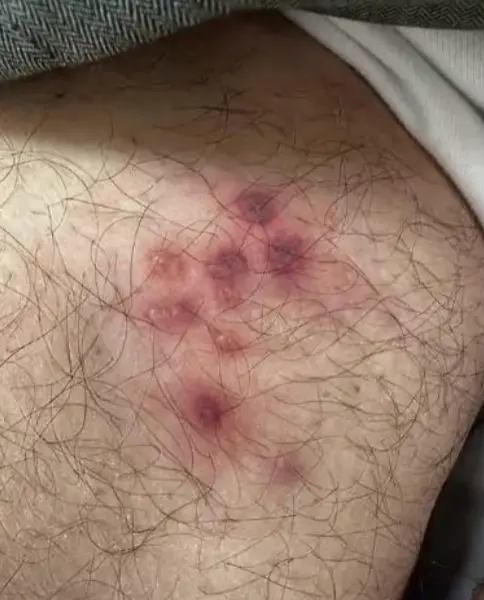
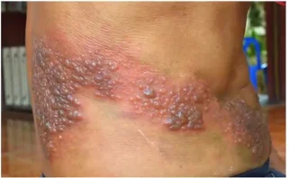
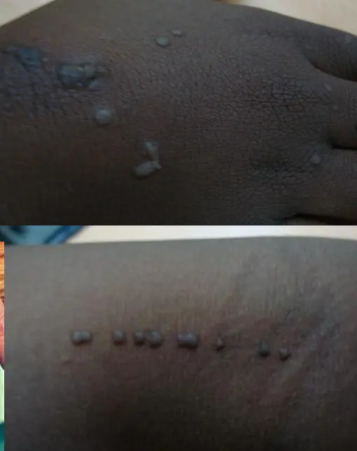
Shingles (Herpes Zoster virus) Z
Context/Case Description:
- Old, pain in flank, Grouped vesicles, pruritic.
- 65 year old man developed a band-like eruption on the left side of his chest. The lesion started as erythematous painful vesicle followed by papule that rapidly blistered. They first appeared in the back and expanded anteriorly till the mid-line.
Diagnosis:
- Shingles / Herpes Zoster
Dermatological picture:
- Erythematous, group of vesicles, dermatomal distribution, mainly in thoracolumbar.
- Band-like eruption on the left side of the chest. Lesions consist of grouped vesicles that started as painful erythema in the area followed by papules that rapidly blistered. They first appeared in the back and extended anteriorly till the mid-line.
Causative organism:
- Varicella-zoster virus (VZV)
Primary or secondary infection? Explain how the disease became active/Explain the course of the disease how it develops:
- Secondary (reactivation) to chickenpox.
- VZV found in Anterior/Posterior Root ganglia (patients with previous chickenpox), where it remains inactive. When immunity decreases, the virus reactivates again, transports anterograde along the sensory nerve, and infects the skin as a secondary infection.
Treatment:
- Acyclovir (most effective if given within 72 hours, though less effective, can still be used after 72 hours).
- Painkiller
- Gabapentin (for Post-herpetic Neuralgia)
- Vitamin B12 complex
Most probable/possible complication:
- Post-herpetic neuralgia (PHN)
Transmission (Chickenpox connection):
- Chickenpox is airborne and infectious before appearance of eruptions.
- If a person who never developed chickenpox (e.g., grandson) comes into contact with a shingles patient, he might develop chickenpox.
Can be a red flag if bilateral or facial involvement
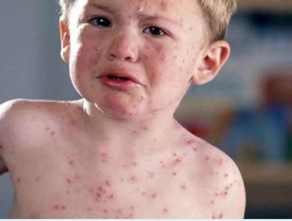
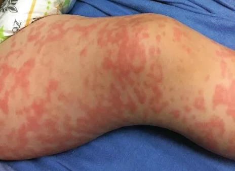
Chickenpox (Varicella)
Context/Case Description & Dermatological picture:
- A 4-year boy developed a generalized vesicular eruption preceded by 3 days of fever and malaise; the eruption started from the face spread down to involve the whole body. The boy is restless, scratching the vesicles.
Most appropriate diagnosis:
- Chickenpox
Differential diagnoses:
- Chickenpox
- Molluscum contagiosum
Clinical Presentation:
- Itchy blisters with central punctum.
- On erythematous base
Causative organism:
- Varicella-zoster virus (VZV)
How is the causative organism spread?
- Spread by airborne droplets to infect other persons with an incubation period of 14 days.
Course of the disease regarding the causative organism’s dormancy and reactivation:
- The causative organism remains dormant in the posterior root ganglia of the spinal cord, then after decades may reactivate itself to affect a dermatomal skin segment as a secondary infection by the organism (Shingles).
Treatment:
- Calamine lotion (for pain relief)
- Systemic antibiotic (to prevent secondary infection)
- Antihistamine: sedation.
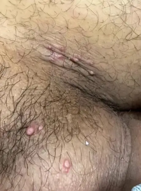
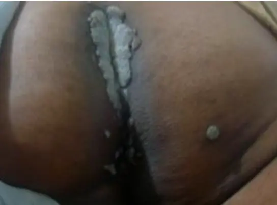 What is the diagnosis?
- Condyloma acuminate / ano-genital warts.
What is the clinical presentation?
- Gray in color.
What is the pathology / causative agent?
- HPV 16, 18.
What are the treatment options?
- Physical destruction:
- Curettage: Warts are anesthetized and then scraped with a curette.
- Cautery: Electrical burning of lesions.
- Cryotherapy: By liquid nitrogen.
- Surgical removal or excision.
- Laser: Is used also for removal of warts.
- Chemical destruction:
- Podophyllin: For mucous membranes, especially genital warts. Should be used cautiously as it is toxic.
- Salicylic acid: In high concentrations.
- Silver nitrate: In form of pencils.