FM
Benign Paroxysmal Positional Vertigo (BPPV)
- BPPV is the most common cause of vertigo, which is a symptom of the condition.
- Though not fully understood, BPPV is thought to arise due to the displacement of otoconia (small crystals of calcium carbonate) from the maculae of the inner ear into the fluid-filled semicircular canals.
- These semicircular canals are sensitive to gravity, and changes in head position can be a trigger for BPPV.
- The posterior canal is the most commonly affected site.
BPV
- Affects an older age group (5th Decade onwards).
- Around 25% of dizziness cases have BPV.
Risk Factors Include:
- Female sex
- Hypertension (HTN)
- Hyperlipidemia
- Cerebrovascular disease
- Menopause
- Allergies
- Migraine
- Chronic obstructive pulmonary disease (COPD)
- Surgical procedure such as a cochlear implant
- Infection
Clinical Features
- Brief severe episodes of vertigo lasting less than a minute.
- The vertigo provoked by sudden changes of head position.
- The reaction decays, so that repeating the movement causes less vertigo.
Other Symptoms of BPPV Include:
- Light headedness
- Loss of balance
- Nausea
- Vomiting
- Nystagmus with positional change of the head
Associated Co-Morbidities
- Meniere’s disease
- Vertebral basilar insufficiency
- Migraine
- Multiple sclerosis
- Infection: sinus or ear
- Thyroid problems
- Reduced bone mineral density
- Sudden hearing loss
Test
- Can be performed in general practice, but usually unnecessary since the diagnosis can be obtained from a careful history.
- Sit the patient on a couch with the head turned to face you.
- Lower the patient, so that his or her head is over the top edge of the couch & 30% below the horizontal.
- Nystagmus provoked by this test is always an abnormal finding.
Nystagmus in BPV
- Is rotatory, beating towards (i.e. Fast phase) the downward ear.
- A latent period of several seconds precedes onset.
- Nystagmus abates after a maximum of 50 seconds (adaptation).
- Violent vertigo accompanies nystagmus.
- On repeating the test, the response diminishes.
Dix-Hallpike Test OSPE
-
To check for right side involvement, rotate the patient’s head to the right 45 degrees while in the long sitting position (this aligns the right posterior semicircular canal with the sagittal plane of the body).
-
The examiner grasps the patient’s head and quickly moves the patient to the supine position with the neck slightly extended (ear down position).
-
The examiner checks for nystagmus. If present, note the latency, duration, and direction (should not last more than 1 minute).
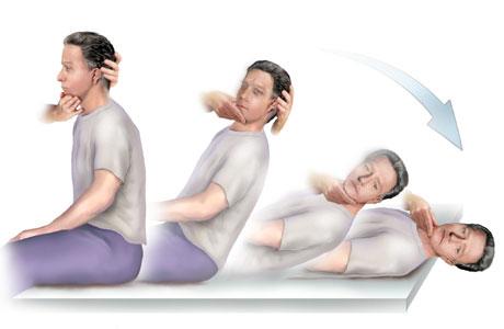
Dix-Hallpike Test
![[Benign Paroxysmal Positional Vertigo BPPV-1741768318273.webp]]
Hallpike Test
Patient Instructions for the Epley Maneuver
- 1. For the next 2 days and nights, keep your head completely vertical.
- To sleep, you might sit in a recliner chair, but do not lie all the way back; just far enough to support your head.
- Support the head by purchasing a neck brace or by pinning two pillows.
- Avoid any head movements upward or downward for the next week.
- Do not sleep on the side that generates your dizziness for an additional week.
- If your right ear is causing the problem, then sleep on your left side.
BPV
- The symptoms persist for an average of 6 months, then resolve.
- The underlying cause is the displacement of calcium crystals (otoliths) in the inner ear.
- Head movement makes the crystals move about, inducing the symptoms.
Treatment
-
Explain the benign nature of the condition.
-
Certain exercises may be used as a way of getting the brain to compensate (90% success has been alleged).
-
Prochlorperazine is the commonly prescribed medication. (Common SE is dystonia).
Epley’s Maneuver
-
Patient starts in long sitting with the head rotated 45 degrees to the affected side.
-
Patient next rapidly reclined to the supine position with the neck slightly extended. This position is held for 30 seconds, or until nystagmus and dizziness subside.
-
The patient’s head is rotated 90 degrees to the opposite side. This position is held for 20 seconds, or until nystagmus and dizziness subside.
-
The patient’s head is turned another 90 degrees, requiring the patient to go from the supine to side-lying position. This position is held for 20 seconds, or until dizziness and nystagmus subside.
-
The patient is brought up to the short-sitting position.
Contraindications to Epley’s Manoeuvre
- Severe carotid stenosis
- Unstable heart disease
- Severe neck disease (cervical spondylosis with myelopathy)
- Advanced rheumatoid arthritis
![[Benign Paroxysmal Positional Vertigo BPPV-1741768358441.webp]]
ENT
Benign positional vertigo develops when crystals of calcium carbonate moves free from Utricle’s glycoprotein membrane and floats within the tube of SCC’s.
Description
- Most common peripheral vestibular disorder.
- Causes: spontaneous, post-traumatic, post-viral infection.
- Affects posterior (90%), horizontal (10%), or superior (rare) semicircular canal.
Presentations
- Recurrent episodes of brief positional vertigo, nausea, and prolonged lightheadedness.
- No hearing loss.
Management
- Typically self-limiting, may recur.
- Education, reassurance, observation.
- Repositioning Procedure (Epley Maneuver-Brandt-Daroff).
- Surgery.
Dix Hallpike Maneuver
-
Used to provoke nystagmus and vertigo commonly associated with BPPV
-
The patient is seated on the exam table with head turned 45 degrees to the side and brought rapidly into the supine position with head hanging over the table. Head supported and rapidly placed into head hanging position.
-
This maneuver allows maximal stimulation of the posterior SCC.
-
Frenzel glasses eliminate visual fixation suppression of response
-
-20250119093947202.webp)