Various changes can be seen on CT and MRI in elderly patients that often bear little correlation with the clinical state of the patient.
Supportive findings:
- Signs of generalized or focal cerebral atrophy
- Enlarged ventricles (ventriculomegaly)
- Narrowing of gyri
- Prominent cerebral sulci (hydrocephalus ex vacuo)
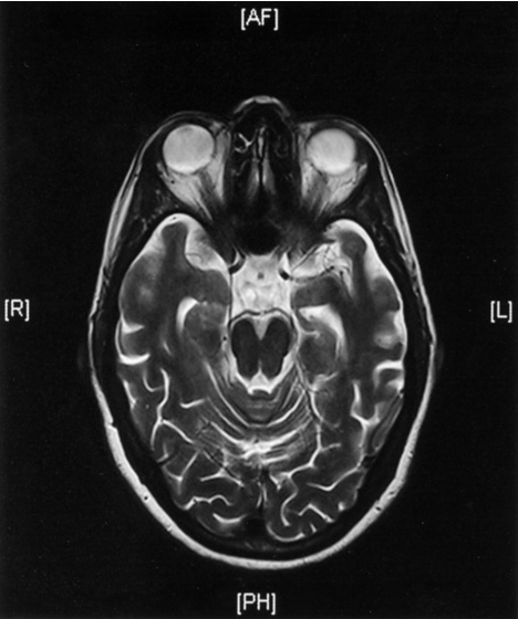
MRI head (T2-weighted; axial plane)
- of a patient with increasing memory disturbance
- Medial temporal lobe atrophy is accompanied by widening of the subarachnoid space and dilatation of the temporal horns of the lateral ventricles
- These changes have been observed in the setting of Alzheimer disease.

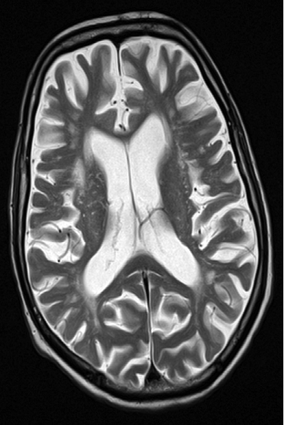
Cerebral atrophy - Axial T2-weighted MRI
showing prominence of the ventricles and generalized widening of the cerebral sulci in keeping with age-related atrophy.
