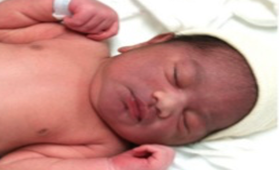
The most common form of traumatic birth injuries are soft-tissue injuries including bruising, petechiae, subcutaneous fat necrosis, and lacerations.
-
Bruising and Petechiae
- Usually self-limiting and are often seen on the presenting portion of the newborn’s body.
- Bruising and edema of the genitals are common findings in infants delivered from the breech position.
- Petechiae of the head and face are often seen in infants delivered from the vertex position, especially with a face presentation.
- Most often, petechiae are present at birth, do not progress, and are not associated with other bleeding.
- Significant bruising has been recognized as a major risk factor for the development of severe hyperbilirubinemia.
- Follow-up within two days of the newborn hospital discharge is recommended for infants with significant bruising to assess them for progressive jaundice.
-
Subcutaneous Fat Necrosis
- Subcutaneous fat necrosis (SFN) is uncommon.
- Usually occurs in the first few weeks of life due to ischemia to the adipose tissue following a traumatic delivery.
- SFN is characterized by firm, indurated nodules and plaques on the back, buttocks, thighs, forearms, and cheeks.
- Typically, this condition is self-limiting.
-
Lacerations and Abrasions
- To the skin from scalp electrodes applied during labor or from accidental scalpel incision at cesarean section.
- Fetal laceration has been reported as the most common birth injury associated with cesarean delivery.
- The lacerations occurred most often on the presenting part of the fetus, typically the scalp and face.
- The majority of fetal lacerations were mild, requiring repair with sterile strips only.
Facial Bruising
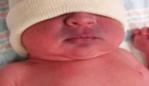
 It is more common when there is a tight nuchal chord, when the delivery is precipitous or difficult, or when the infant is bundled. This facial appearance could be mistaken for cyanosis, but with a quick comparison to the color of the rest of the body, the diagnosis is obvious.
This type of bruising resolves over the course of several days.
Perioral Cyanosis

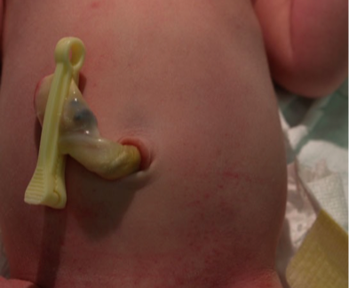
Petechiae of Newborn
 While petechiae may be due to pressure during birth, widespread petechiae deserve some evaluation.
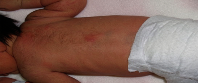
Subcutaneous Fat Necrosis
- This red lesion is subcutaneous fat necrosis.
- On palpation, there is a firm nodule in the subcutaneous tissue under the area of redness that is freely mobile with respect to the bony structures underneath it.
- Subcutaneous fat necrosis is more common in infants who have had difficult deliveries, cold stress, or perinatal asphyxia.
- Lesions are typically asymptomatic and resolve spontaneously within several weeks, usually without scarring or atrophy.
- Infants with extensive lesions or with renal disease should have calcium levels followed once or twice weekly.
- Hypercalcemia associated with subcutaneous fat necrosis is rare, but is a potentially lethal complication
