Chest X-ray Indications:
All patients suspected of having current Pneumonia
What structures can we see?
LUNGS, MEDIASTINUM, BONY CAGE, SOFT TISSUE COMPONENT
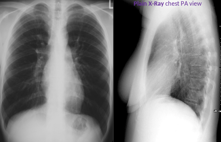
- Lungs are equally Lucent - central opacities in lung are majorly bronchial arteries - area is called hilum ; concave shaped, left is higher; abnormal if lobulated
- Lung becomes more luscent from upward to downward lateral view Z
- Heart slightly deviated to left
- Aortic Arch
- Assessment of heart size; should be less than half of width of thoracic ((CC))
- Assess heart borders differentiating density between air and tissue
- Assess Tracheal Deviation; should be central
- Right diaphragm should be higher than left; hepatomegaly, pneuomo-perotineum
- Costrophrenic angle sharp on left central side of diaphragm Z
- Assess bones structures: Clavicles, Ribs, Scapula, Spine
- Soft Tissue assessment

X-ray chest (PA view)
Lungs : The lung volumes are normal. No abnormal pulmonary parenchymal opacities are seen.
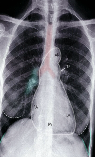
Heart : The cardiac silhouette (outlined in white).
Hilum : Pulmonary hila (example indicated by green overlay).
Mediastinum :Superior mediastinum are normal in size and configuration.
Costophrenic angles: The lateral costophrenic sulci (outlined in green) are sharp, with no evidence of effusion.
Bone & soft tissue : No concerning skeletal lesions are identified.
LV: left ventricle; RV: right ventricle; RA: right atrium; PT: pulmonary trunk (white arrow); AK: aortic knob; Trachea: red overlay, Breast shadow: white dotted outline
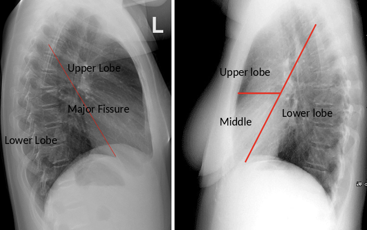
Fissures and lobes
