IM
DR WAQAR
OBJECTIVES
By the end of this lecture, student should know:
- Difference b/w PUD & dyspepsia
- Etiologies
- Signs and Symptoms
- Investigation for H. Pylori and peptic ulcer
- Treatment of Peptic ulcer
- Know which tests to do after treatment
- What is Zollinger Ellison Syndrome
CASE 1
25 year old male Pain in epigastric area, off and on since 2 months. Relieved with food Smoker, on NSAIDS since long time Very stressful job Physical examination normal Name some differentials
Case 2
50 year old female Epigastric pain since 3 months off and on. More after eating Pale stools which are difficult to flush Weight loss of 10 kg in 3 months Chronic alcoholic. Serum triglycerides very high Jaundice present What is your differential?
Dyspepsia
What is Dyspepsia ?
The term “ dyspepsia” is a broad term and includes any of the following:
- Heart burn
- Food related discomfort in the upper abdomen
- Nausea, gas etc
- Feeling of fullness after eating a little
Every dyspepsia patient does not need urgent & detailed investigations. However, presence of any of the following warrants urgent workup (alarm symptoms)
- Dysphagia
- Weight loss
- Severe Vomiting
- Severe anorexia
- Hematemesis
- Melena
PEPTIC ULCER
Definition:
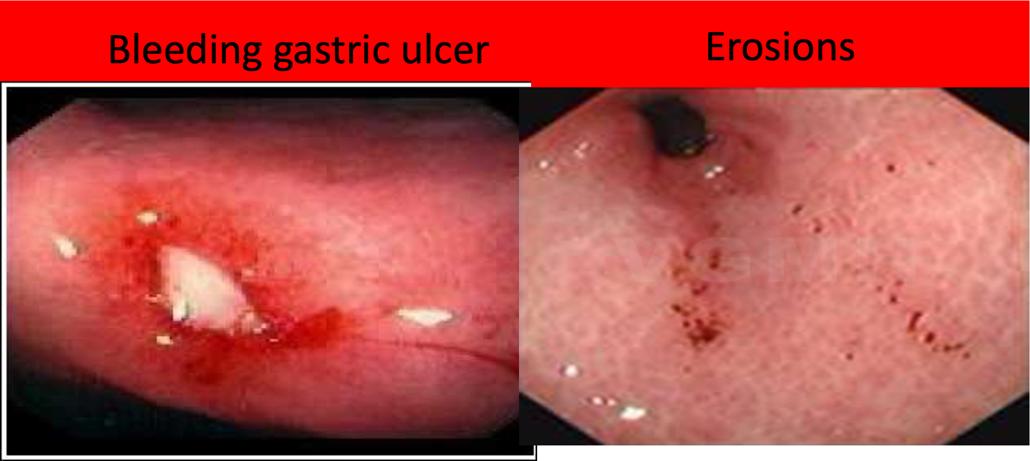
It is a break in the gastric /duodenal mucosa, extending down to the muscularis layer.
EROSIONS: Superficial breaks in the mucosa alone, not extending deep.
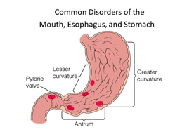
Most common sites of peptic ulcers:
- Duodenal bulb (most common)
- Lesser curvature
- Antrum of the stomach
SOME GENERAL POINTS
| Feature | Duodenal Ulcers | Gastric ulcers |
|---|---|---|
| Frequency | Much more common than gastric ulcers | Less common |
| Location | Usually in the duod. bulb(1st part) | |
| Malignancy | Almost never malignant | May be malignant |
| Pain with eating | Eating relieves pain | Eating worsens the pain |
| Age | Typically younger | Usually above age 50 |
ETIOLOGY OF ULCERS
- Helicobacter Pylori infection -⇒ Most Common
- NSAID/ Aspirin use ⇒ Most Common
- Hyperacidity states (eg Zollinger Ellison syndrome)
- Biphosphonates ( medicines used in osteoporosis)
- Stress
- Alcohol, dietary factors & smoking may aggravate the symptoms.
SO: In patients with peptic ulcers, the first 2 things to rule out are:
- H. Pylori infection (by tests)
- NSAID use (by history)
Some points about H. Pylori
- It is a bacterium infecting the gastric & duodenal mucosa
- 2/3rd of the world population is infected
- Poor hygienic conditions → more chances of infection
- Can cause “no” symptoms OR erosions, gastritis, ulcers
***Points about ulcer producing drugs ***
- Long term use of aspirin & ‘traditional’ NSAIDs (diclofenac, ibuprofen etc), increases the risk of peptic ulcers. ( gastric more than duodenal)
- Use of these meds in ulcer patients → high risk of complications
- COX 2 inhibitors(which are also NSAIDs) have much less risk of peptic ulcer disease ( eg celecoxib). They may be called “gastro-protective” )
- Long term steroid use increases ulcer risk
So, ulcer producing drugs are:
- NSAIDs
- Aspirin
- Steroids
SIGNS & SYMPTOMS of ULCERS
-
Asymptomatic: 70% of patients can be totally asymptomatic, specially those on NSAIDs.
-
Epigastric pain:
- Classic symptom
- Usually a dull ache
- Relieved by antacids
- Food relieves pain in DU
-
Nausea, belching, heartburn, bloating etc. ( dyspeptic symps).
-
Physical exam is usually normal ( may be mild epig. tenderness)
Clinical S/S can not distinguish between DU & gastric ulcer
Complications of PUD
- Bleeding (most common complication)
- Perforation (can lead to ? )
- Ulcer in the pylorus can become fibrosed/scarred → gastric outlet obstruction → persistent vomiting
ZOLLINGER ELLISON SYNDROME
It is a syndrome due to a gastrin secreting tumor (usually in the pancreas or duodenum).
Excess gastrin → hyperacidity → duodenal ulcers & diarrhea
When to suspect:
- Ulcers not responding to usual treatment
- ***Large ulcer ***
- Multiple ulcers
DIAGNOSIS:
- Fasting serum gastrin levels : High
- Serum gastrin levels after giving i.v. “secretin” ( secretin stimulation test). In gastrinoma, further rise in serum gastrin levels
Rx: Surgical removal of the tumour
RAPID FIRE QUESTIONS
-
Difference between ulcer & erosion? ulcer is deeper; superficial
-
Name 2 most common causes of peptic ulcer? Nsaids, h pylori
-
Which medicines can cause ulcer? biphosphatase nsaids
-
Name 4 alarm symptoms in dyspepsia patient, where endoscopy is LAAZIM? hematemisis, melena, weightloss, vomitting
-
Name 3 most common areas where ulcers occur? duodenum pul, epigastric, pylorus
-
Food worsens the pain in which ulcer? gastric when you eat food, duodenal better
-
Which ulcers can be malignant, duod or gastric? commonly in gastric
-
Symptoms suggesting ulcer disease. What are the next 2 steps to do? Endoscopy, H. Pyolri testing
-
H. Pylori can cause what? gastritis, Ulcer gastric/duodenal, asymptomatic
-
Which NSAIDs are gastro-protective? ?
-
Can ulcer patients be asymptomatic? yes
-
Name the 4 tests for H. Pylori? Which is NOT the best? Urea breath test, serum anti body titers, …, …
-
Which enzyme is produced by H. Pylori? urease
-
Patient is taking PPI form before. You want to do H.Pylori test. Whats the first step to do? 2 weeks stoppage before testing
-
What are the “symptomatic” treatments for peptic ulcer disease? antiacids, PPI, Bismo
-
Which drug was withdrawn in the U.S? tidine..
-
Treatment of H. Pylori is “3 drugs for 10 days”, right or wrong? wrong - 4 drugs for 2 weeks
-
Treatment of H. Pylori? Name any 1 regimen? choclate…
-
After treatment, which test to do to confirm eradication? When to do? Urea breath test - after month
-
When to give prophylactic PPIs? Name any 2 situations? Nsaids
-
3 complications of ulcer disease? Perforation, malabsorption
-
What is high in Zollinger Ellison syndrome? gastrin secretin
-
When to suspect this syndrome? large ulcer, multiple
-
Rx of Zollinger? Surgery
SURGE
- Peptic ulcers are found approximately four times as often in the duodenum than in the stomach
- Duodenal ulcers are more common in men
- Gastric ulcers equally affect men and women
- Ulcers are found at all ages
- More common with increasing age
- Whilst acid attack is a marked feature of duodenal ulcer (DU),
- impaired mucosal resistance appears to be more important for gastric ulcer.
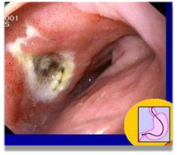
DUODENAL ULCER: Risk Factors:
-
Cigarette smoking, stress, NSAIDs, corticosteroids and infection with H. Pylori
-
NSAIDs—Deplete endogenous mucosal prostaglandins making the mucosa more susceptible to ulceration.
-
Smoking—Increases acid secretion, impairs ulcer healing, accelerates gastric emptying and decreases pancreatic bicarbonate secretion.
-
H. pylori—Over 90 percent patients with a duodenal ulcer have this organism
Clinical Features: DU
- Epigastric pain—Most frequent symptom, appears in empty stomach and relieved by taking food.
- Appetite is good.
- Malignancy almost never occurs.
- Vomiting not so common.
- Periodicity comes and goes in a 4 to 6 months cycle.
- Duration of attack—A month or two.
Peptic ulcer perforation
‘Boardlike rigidity’ of diffuse peritonitis However, in the elderly and the critically ill presentation can be atypical. Subdiaphragmatic free gas on a plain erect chest X-ray is diagnostic. The availability of Helicobacter pylori eradication regimens and potent proton-pump inhibitors alleviate patients’ ulcer diathesis without the need for a definitive acid-reducing procedure. A simple omental patch repair and thorough peritoneal toileting to be done during the emergency operation. The only exception is giant duodenal perforations (>2 cm), not be amenable to a simple patch repair
Z OSPE EX
 Erect chest radiograph showing right subphrenic free gas shadow associated with a perforated peptic ulcer.
A small perforation at the juxtapyloric area
Pedicle omental patch (Graham) repair on the perforation site secured with absorbable sutures
GASTRIC ULCER
Risk Factors :
-
Tobacco, alcohol and H. pylori infection.
-
10 percent of people taking NSAIDs, suffer from acute gastric ulcer.
-
Gastric ulcer frequently develops in degenerate and aged gastric mucosa.
Site Gastric ulcer is most frequently seen near the incisura, a constant notch in the lower part of lesser curvature.
Age/Sex Gastric ulcers occur most commonly in elderly women with the highest incidence between 55 and 60 years of age.
Clinical Features:
-
Pain occurs soon (15–30 min) after eating, which is the precipitating factor unlike duodenal ulcer.
-
Relieving factor—vomiting.
-
There may be loss of weight and hematemesis is more common than melena.
-
Diagnosis by endoscopy and biopsy.
A good clinical rule is to suspect initially that every gastric ulcer is malignant.
FM
Peptic Ulcer Disease (PUD)
Overview
- Refers to an ulcer in the lower oesophagus, stomach, or duodenum.
- Ulcers in the stomach or duodenum may be acute or chronic; both penetrate the muscularis mucosae but the acute ulcer shows no evidence of fibrosis.
- Erosions do not penetrate the muscularis mucosae.
The term ‘peptic ulcer’ refers to an ulcer in the lower oesophagus, stomach or duodenum, in the jejunum after surgical anastomosis to the stomach or, rarely, in the ileum adjacent to a Meckel’s diverticulum.
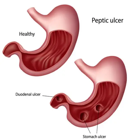
Risk Factors for PUD
- Less than before
- P/H ulcer, recurrence more likely
- Risk factors include:
- H-pylori
- Family history
- NSAID use
- Cigarette smoking
- Chronic renal failure
- Blood group “O”
Diagnostic Difficulties
- Not textbook presentation
- Early presentation
- History:
- ALARM symptoms ??
- Specific symptoms
- NUD
- MI ??
- NSAID
- Smoking
Red Flags of Dyspepsia
- Unintended weight loss
- Severe vomiting
- Progressive dysphagia
- Odynophagia
- Unexplained or iron deficiency anemia
- Hematemesis
- Jaundice
- Palpable abdominal mass
- Lymphadenopathy
- Family history of upper GI cancer

Odynophagia (from the Greek roots odyno-, pain + -phagia, from phagein, to eat) is painful swallowing, in the mouth (raise suspicion of gastric malignancy):
Alarm Symptoms
- Anorexia
- Loss of weight (progressive & unintentional)
- Anaemia due to iron deficiency
- Recent onset of persistent symptoms: vomiting
- Melaena, haematemesis
- Dysphagia (progressive)
- Epigastric mass or
- Suspicious barium meal.
Helicobacter Pylori (H. pylori) Bacteria
-
H. pylori is a slow-growing spiral Gram-negative flagellate urease-producing bacterium which plays a major role in gastritis and peptic ulcer disease.
-
It colonizes the mucous layer in the gastric antrum.
-
But is found in the duodenum only in areas of gastric metaplasia.

H. Pylori Overview
- Gram –ve flagellated spiral
- Casually related to:
- GU
- DU
- Gastritis
- Gastric B – cell lymphoma
- Gastric adenoma
- Prevalence - high
- More in developing countries
- Roughly related to age
- Saudi local study 67-89%
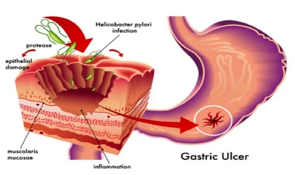
Therapeutic
Peptic Ulcer Definition
ulcer which occur in presence of acid and pepsin (penetrate muscularis mucosa). It is defined as a chronic inflammatory condition characterized by ulceration in regions of the upper gastrointestinal tract where parietal cells secrete pepsin and hydrochloric acid. The most common sites are the duodenum and stomach.
An ulcer of the alimentary tract mucosa, usually in the stomach or duodenum, & rarely in the lower esophagus, where the mucosa is exposed to the acid gastric secretion
It has to be deep enough to penetrate the muscularis mucosa
Other types Definitions: Ulcers Erosion
Types:
1-Acute : ( superficial gastric erosions): due to
- ulcerogenic drugs e.g NSAID and aspirin
- Stress ulcer e.g MI, hypotension due to hemorrhage….etc.
2-chronic ulcer
Mode of transmission:
Transmission is from person to person, and an important mode of spread may be gastro-oral (ie, through exposure to vomitus or saliva).
Epidemiology:
H.pylori infection is commoner in developing countries and is associated with low socioeconomic standards worldwide. The majority of infections are probably acquired in childhood.
Etiology:
Major causes of peptic ulcer: The majority of PUD patients are H. pylori infected
- H. pylori infection (previously discussed) ( 70-80%)
- Aspirin and other Non steroidal anti-inflammatory drugs (NSAIDs). Most of drug induced ulcers are dose related. They decrease mucosal resistance (main factor in GU
- Acid hypersecretory states such as Zollinger-Ellison syndrome. (gastrin hormone production)
- Up to one-fourth of ulcers are idiopathic.
- excess vagal stimulation (stress and anxiety)
- decreased gastrin and histamine destruction as in liver cell failure and hepatic cirrhosis
- allergic conditions (histamine)
N.B.: Z Cyclooxygenase-2 (COX-2) inhibitors: Celecoxib, rofecoxib, and valdecoxibe are NSAIDs that selectively inhibit cyclooxygenase-2 (COX-2)-the principal enzyme involved in prostaglandin production at sites of inflammation-while sparing cyclooxygenase-1 (COX-1), the principal enzyme involved in the synthesis of prostaglandins important in gastric cytoprotection.
Regulation of HCl secretion
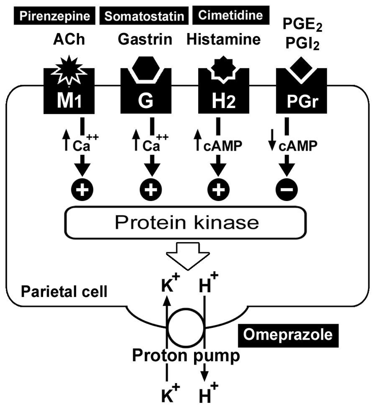 Best general drug is proton pump inhibitor - Omeprazole
Clinical sequelae:
-
Although chronic H pylori infection with gastritis is present in 30-50% of the population, the majority are asymptomatic and suffer no sequel.
-
Peptic ulcer disease (especially duodenal ulcer); however, only J 5% of people with chronic infection develop a peptic ulcer.
-
Chronic H pylori gastritis
-
Increased risk of gastric adenocarcinoma and low-grade B cell gastric lymphoma (mucosa-associated lymphoid tissue lymphoma; MALToma).
Clinical picture of peptic ulcer:
(A) Typical presentations: Symptoms: Patients can be asymptomatic or have anorexia, nausea, vomiting, heartburn or epigastric pain.
Ulcer pain or ulcer dyspepsia:
| Feature | DU (Duodenal Ulcer) | GU (Gastric Ulcer) |
|---|---|---|
| Etiology | Contact of acid with exposed nerve endings in the base of the ulcer | Contact of acid with exposed nerve endings in the base of the ulcer |
| Character | Deep seated burning sensation (may be dull, colicky, or stabbing) | Deep seated burning sensation (may be dull, colicky, or stabbing) |
| Time | pain occur 0 .5- 2 hours after meal (hunger pain) to buffer hyperacidity. Also occur at night (nocturnal pain which awake patient at midnight) | 0.5-1 hour after meal |
| Site | Epigastric pain to the right of middle line | Epigastric pain to the left of middle line |
| Factors that increase pain | Hunger, irritant foods, smoking, stress, tea, coffee, drugs | Food, … (other factors TBD) |
| Factors that decrease pain | Alkalies, eating | Alkalies, vomiting |
| Signs | Localized tenderness at the site of pain (right of middle line) | Localized tenderness at the site of pain (left of middle line) |
| Appetite | Weight gain | Weight loss |
(B) Atypical presentation:
- Painless ulcer: may be (bleeding, perforation, DM….etc
- Complicated ulcer: 1- GIT bleeding: may be —Occult blood in the stool -severe hemorrhage with haematemesis and melena 2- Perforation 3- Malignancy
The differences between DU and GU can be slight, making differentiation difficult based on symptoms alone so, the accurate diagnosis depends on radiological (barium meal) or endoscopic visualization of the ulcer.
Physical examination
It is often normal in uncomplicated peptic ulcer disease. Localized epigastric tenderness to deep palpation may be present. Pointing tenderness in the epigastrium in D.U.
Signs of complication:
- Pallor due to anemia due to chronic blood loss.
- Haematemsis and melena.
- Gastric carcinoma in gastric ulcer.
Pathophysiology of peptic ulcer :
Normally there is balance between : 1) Defensive mechanism in the stomach due to
- mucosa secrete mucous and bicarbonate
- mucosa rapidly replaces damaged epithelial cells
- mucosa synthesizes PGE1 and PGE2 which (stimulate synthesis and secretion of mucous and bicarbonate and increases gastric blood flow)
So, mucous and bicarbonate form protective gel like over the mucosa which with the epithelial cells tight junction form mucosal barrier which prevent (back diffusion of acid (H) , also Na and K diffusion to gastric mucosa)
2)Aggressive factors in the stomach:
-
secretion of HCL and pepsin(proteolytic enzyme)
-
infection of the stomach specially in the duodenum with helicopacter pylori (G-ve bacilli)
-
Ulcers extend through the muscularis mucosae and are usually over 5 mm in diameter. Ulcers
-
occur five times more common in the duodenum than the stomach. Over 95% of the duodenal
-
ulcers are in the bulb or pyloric channel.
Incidence: Peptic ulcer disease (PUD) is a common disorder that affects millions of individuals worldwide
Although ulcers can occur in any age group, duodenal ulcers most commonly occur between the ages of 30 and 55, whereas gastric ulcers are more common between the ages of 55 and 70.
Complications of peptic ulcer:
- Hemorrhage: Is the most common complication, it is an emergency situation and is lethal in I 0% of patients, the mortality increases with age. It presents with anemia with occult blood in stools, hematemesis, vomiting of coffee ground material, melena or with shock.
Treatment: The patient needs to be admitted to the ICU, resuscitated by fluids and blood. Endoscopy upper GI to know the cause of the bleeding, should b e done Intravenous proton pump inhibitors.
- Perforation:
-
It usually presents with sudden onset of severe abdominal pain; ileus and board like rigidity of the abdominal wall.
-
The first presentation may be shock and septicemia. Polymorph nuclear leukocytosis and raised serum amylase may be found.
-
Plain X-ray abdomen in the erect position may show air under the diaphragm.
Management: Nasogastric tube to empty the stomach and prevent further peritoneal soiling. IV fluids and broad spectrum antibiotic, followed by surgical closure and omental patch.
-
Penetration: Into the pancreas or the biliary system presents with increasing severity of the pain with radiation to the back and symptoms and signs of pancreatitis and cholangitis.
-
Gastric outlet obstruction:
- It presents with epigastric fullness, vomiting of foul odor of previously ingested food with alkalosis , loss of weight is seen in longstanding cases.
- The obstruction may be due to edema and inflammatory reaction around the ulcer.
- Barium meal and endoscopy are diagnostic.
Management: Naso-gastric suction for one week combined with IV fluid replacement and proton pump inhibitors. The most common cause is fibrosis and scarring and this needs endoscopic balloon dilatation or surgery.
- Malignant change in gastric ulcer