Main Salivary Glands
-
There are 3 pairs of main salivary glands: - Parotid Gland - Submandibular Gland - Sublingual Glands
-
Plus small innumerable glands
- scattered in the mucous membrane of the oral cavity.
- scattered in the mucous membrane of the oral cavity.
Minor Salivary Glands
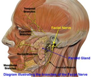
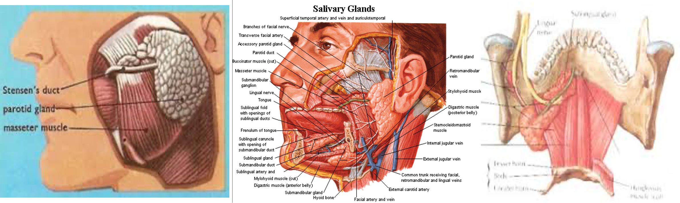
Parotid Gland
-
Serous cells only.
-
On the side of the face, deep to the skin, subcutaneous tissue, superficial to the masseter muscle.
-
Tail of parotid extends superficial to SCM.
-
Stensen’s duct begins at the anterior border of the gland 1.5cm below the zygoma.
-
Traverses the masseter 5-6 cm, pierces the buccinator.
-
Opens in the mouth lateral to the 2nd upper molar.
Submandibular Gland
- Mucous and serous cells.
- Submandibular triangle: between anterior and posterior bellies of digastrics’ and inferior margin of the mandible.
- Medial and inferior to the mandible.
Wharton’s Duct
- Exits the gland from the medial surface.
- Travels between the hyoglossus and mylohyoid muscles.
- Enters the genioglossus muscle.
- Opens into the mouth just lateral to the lingual frenulum.
- CN XII is inferior to the duct and the lingual nerve is superior to the duct.
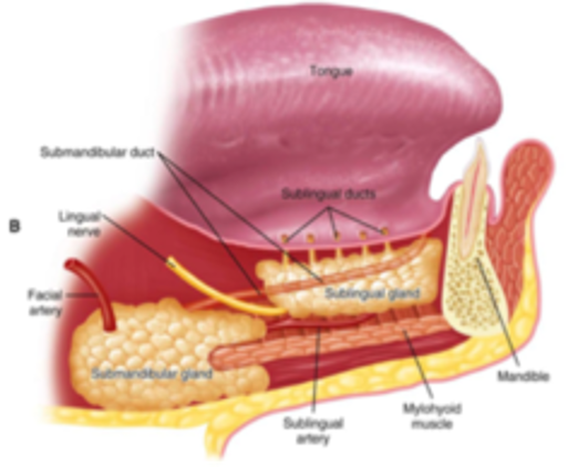
Sublingual Glands
- Mucous secreting.
- Just below the floor of the mouth mucosa.
- Bordered by genioglossus/hyoglossus medially.
- Mandible laterally.
- Mylohyoid inferiorly.
- Wharton’s duct and lingual n. travel between the SL gland and genioglossus muscle.
- No fascial capsule.
Ducts of Rivinus
- Ducts of Rivinus (~10) along the superior aspect of the gland.
- Open into the mouth along the sublingual fold in the floor of the mouth.
Minor Salivary Glands
- Either mucous/serous or both.
- 600-1000/person.
- Each gland has its own duct.
- Found most commonly in the buccal, labial, palatal, and lingual regions.