Neuro-ophthalmology
-
Afferent
-
Efferent
-
Anatomy
-
Examination
-
Diagnoses
-
Tests
Optic Nerve Anatomy
- Cranial nerve II, the second of twelve paired cranial nerves.
- These fibers are axons of the retinal ganglion cells of one retina.
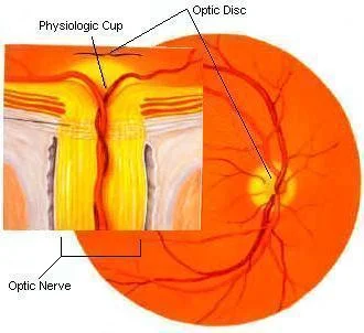
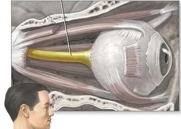
- The optic nerve is ensheathed in all three meningeal layers (dura, arachnoid, and pia mater).
- It leaves the orbit (eye socket) via the optic canal, running postero-medially towards the optic chiasm.
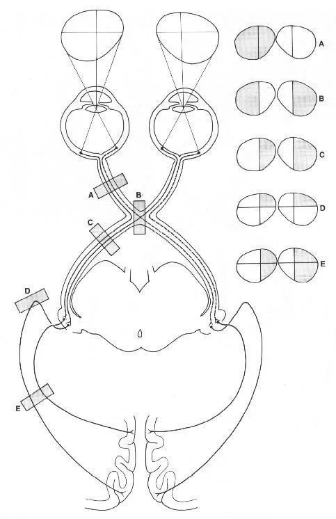
Visual Pathway Diagram
- Eyeball
- Retina
- Optic nerve (II)
- Optic chiasma
- Optic tract
- Lateral geniculate nucleus of thalamus
- Optic radiation
- Visual cortex
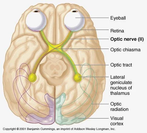
- In optic chiasm there is a partial decussation (crossing) of fibers from the temporal visual fields (the nasal hemi-retina) of both eyes.
- From there, the nerve fibers become the optic tract.
- Passing through the thalamus and turning into the optic radiation.
- Until they reach the visual cortex in the occipital lobe at the back of the brain. This is where the visual center of the brain is located.
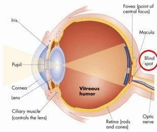
Optic Nerve Physiology
-
The eye’s blind spot is a result of the absence of photoreceptors in the area of the retina where the optic nerve leaves the eye.
-
Fiber tracks of the mammalian central nervous system (as opposed to the peripheral nervous system) are incapable of regeneration, and, hence, optic nerve damage produces irreversible blindness.
Afferent Anatomy
Examination
- Visual acuity
- Color vision
- Visual field
- Pupil examination
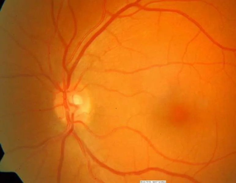
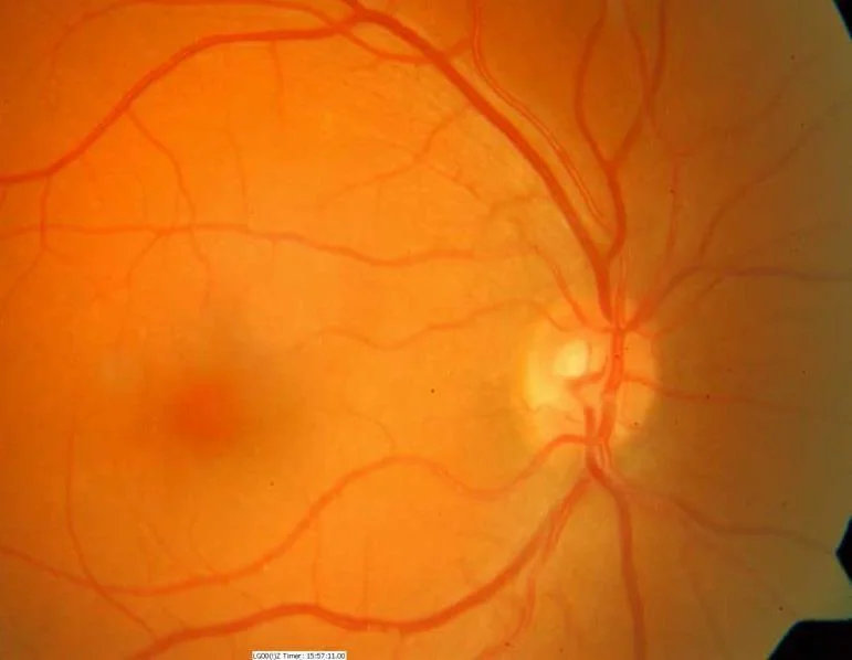
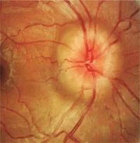
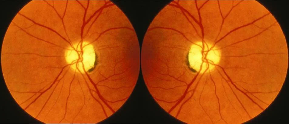
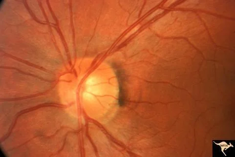
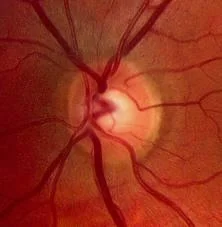
- Fundoscopy
Fundus Exam