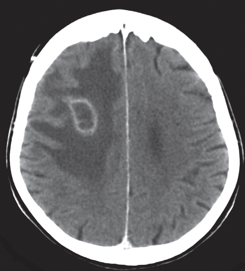
Pus in the center of the abscess appears as low density on CT or fluid on MRI. The wall of the abscess enhances with intravenous contrast and may be surrounded by oedema, giving an appearance known as ‘ring enhancement’
Cerebral abscess.
Post contrast CT scan showing a right frontal ring-enhancing lesion with surrounding vasogenic oedema

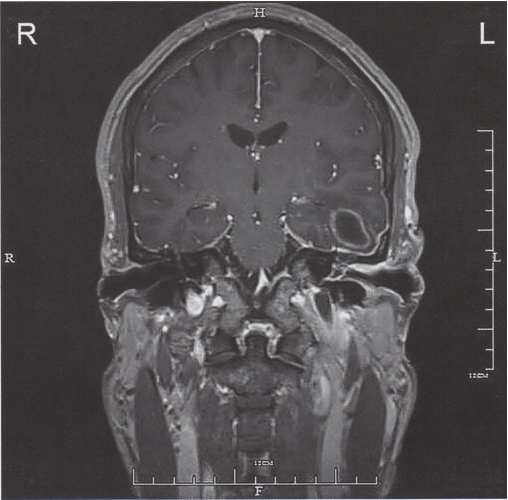
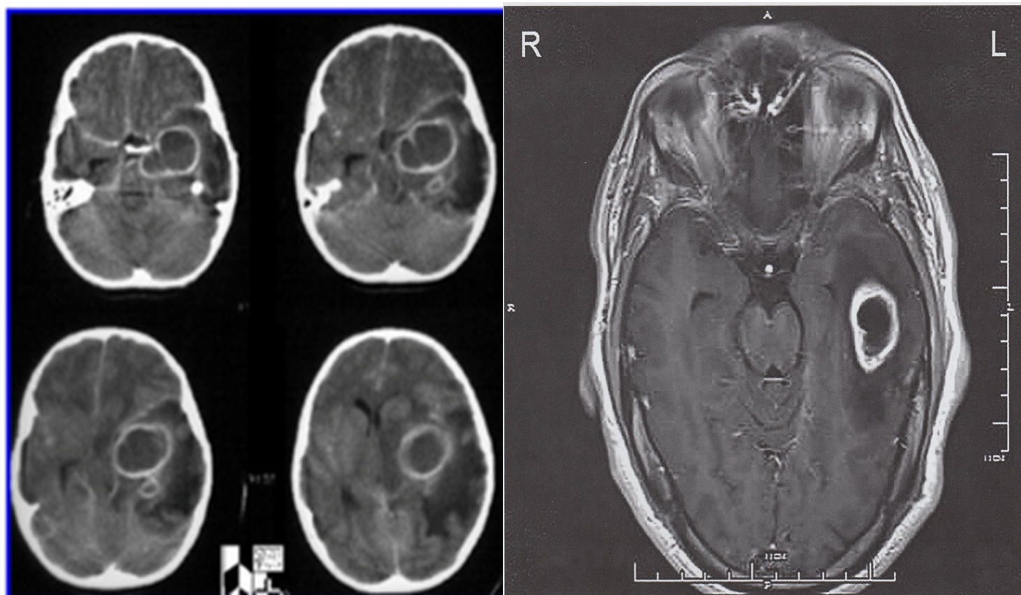
Ring-enhancing lesion left temporal lobe
MRI head (T1 weighted; with contrast; coronal plane) of a patient with a history of left advanced otitis media In the left temporal lobe, there is an oval lesion with ring-like contrast enhancement, central low signal intensity necrosis, and perifocal edema. The walls of the left ear canal and middle ear are thickened and show contrast enhancement consistent with otitis media and otitis externa.
The appearance of the temporal lobe lesion is compatible with an abscess.

Cranial MRI (with contrast, axial view)
An oval lesion can be seen in the left temporal lobe, with ring-like contrast enhancement and central necrosis . The perifocal edema that is visible corresponds to the lesion. Differential diagnoses include an abscess, glioblastoma, and metastasis.
Diagnosis: abscess following advanced otitis media
