IM
MOST IMPORTANT PFT
SPIROMETRY
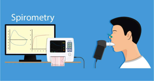
- A spirometer is an apparatus used to measure the volume of air inhaled and exhaled by the lungs.
- After attaching the mouth piece, patient inhales maximally then exhales forcefully. This is recorded by the machine.
Two very important measurements by the spirometer are:
-
FEV1: forced expiratory volume in 1 second.
- This is the amount of air which comes out in
- the 1st second of forced expiration.
-
Vital capacity( also called forced vital capacity or FVC)
Remember: most of the air comes out in the 1st sec. of exhalation
FEV1/ VC
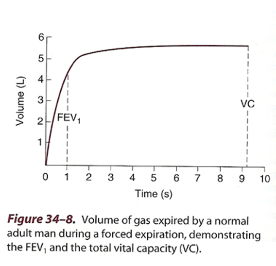 The FEV1 is used to classify the severity of obstructive lung diseases traditionally based on 0% predicted values into five levels.
- FEV1>70% of predicted is mild
- FEV1 60-69% of predicted I is moderate
- FEV1 50-59% of predicted is moderately severe
- FEV1 35-49% of predicted is severe
- FEV1 <35% of predicted is very severe
SOME NORMAL VAUES
- TV: 500 cc
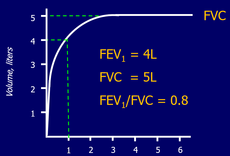
- F.V.C. : 5 liters(5000cc)
- FEV1 : 4 liters (4000cc)
- TLC: 5.7 liters (5700cc)
- FEV1/FVC ratio : 4/5 = 75% to 80%
USES OF SPIROMETRY
- Diagnosis of certain lung diseases eg asthma, COPD, interstitial lung diseases (obstructive pattern vs. restrictive pattern)
- To see the effect of medicines being given for those diseases
- Pre-operatively, to assess lung function
Spirometry results in some lung diseases
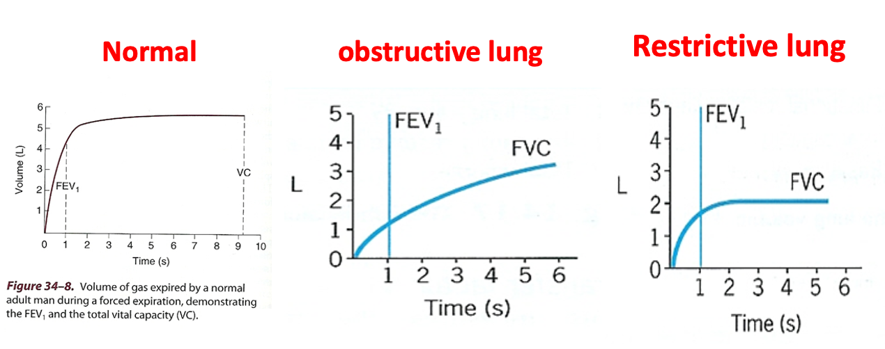
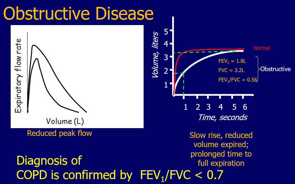
1) Obstructive diseases (asthma, COPD)
- a) FEV1 is too much reduced
- b) FVC is slightly reduced, so
- **c) FEV1 /FVC ratio is
- reduced.**
- **d) TLC is increased
- ( air trapped inside)**
- **eg FEV1= 1.3L, FVC = 3.2L, ratio
- will be (1.3/3.2) x 100: 41%**
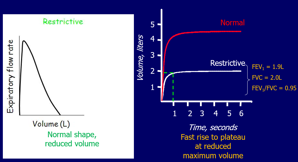
2) Restrictive diseases ( pulm. fibrosis, decreased chest movement)
- a) FEV1 reduced
- b) FVC equally reduced, so
- **c) FEV1/ FVC ratio is normal **
- d) TLC is decreased
- Eg: FEV1 1.7L, FVC 2L, ratio * ( 1.7/2) x100 : 85%
Thera
Is the most common of the Pulmonary Function Tests (PFTs)
Is a method of assessing lung function by measuring the volume(amount)& flow(speed) of air the patient can expel from the lungs after a maximal inspiration
-
Measure airflow obstruction to help make a definitive diagnosis .
-
Distinguish between Obstructive and Restrictive diseases of the lungs.
Types of Spirometers
| Parameter | Description |
|---|---|
| Bellows spirometers | Measure volume; mainly in lung function units |
| Electronic desk top spirometers | Measure flow and volume with real time display |
| Small hand-held spirometers | Inexpensive and quick to use |
 | |
| pneumotachometer (1st) | |
| Electric Spirometry (3rd) Plethysmograph; | |
| measures DLCO (5th) |
Pulmonary function test
Lung volumes and capacities
Standard Spirometry Indices
The Spirometer calculates different ventilation parameters:
| Parameter | Description |
|---|---|
| FVC - Forced vital capacity | The total volume of air that can be forcibly exhaled in one breath |
| FEV1 - Forced expiratory volume in one second | The volume of air expired in the first second of the blow |
| FEV1/FVC ratio | The fraction of air exhaled in the first second relative to the total volume exhaled |
Predicted Normal Values Based on Following
- Age
- Height
- Weight
- Sex
- Ethnic Origin calculated number will be given to estimate viability
Volume-time loop
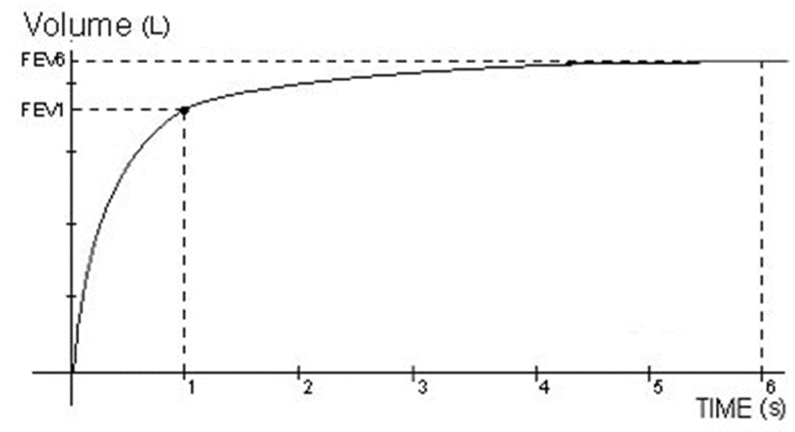 The volume versus time curve is a an alternative way of plotting spirometric results and is another useful illustration of patient performance.
Normally the whole FVC is expelled in four seconds 80% in second normally
| Parameter | Description |
|---|---|
| Forced expiratory volume (FEV1) | the volume of air expelled in the first second of a forced exhalation. In normal subjects 75-80% of the FVC can be expelled in the first second. |
| FEV1/FVC | the normal value is 75-80%. Anything below this is considered abnormal (obstructive or restrictive disease). |
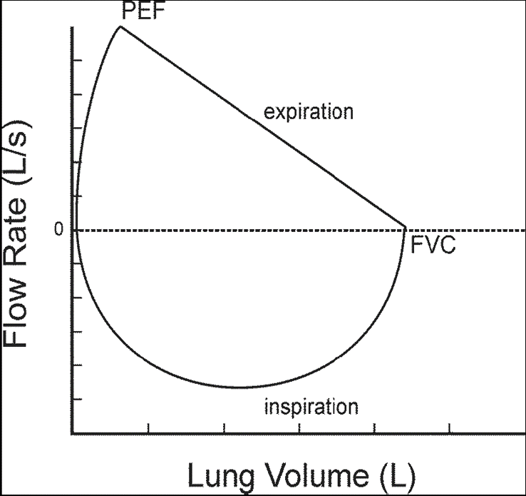
Flow- volume loop Y
-
Spirometry is a valuable tool for analyzing the flow rate of air passing into and out of the lungs.
-
Flow volume loops provide a graphical illustration of a patient’s spirometric efforts.
Spirogram Patterns
- Normal
- Obstructive
- Restrictive
- Mixed Obstructive and Restrictive Y
obstructive and restrictive diseases Z
Lung disease is often divided into two broad categories: obstructive disease and restrictive disease.
-
Examples of obstructive disease
are Emphysema, Chronic Bronchitis, and bronchial Asthma. -
Examples of restrictive diseass are abnormalities of the spine and chest and diseases within the lungs that make them less elastic (“stiffer”), such as pulmonary fibrosis.
| Criteria | FEV1: % predicted | FVC: % predicted | FEV1/FVC |
|---|---|---|---|
| Normal | > 80% | > 80% | > 0.7 |
| Obstructive Disease | < 80% ↓↓ | < 80% ↓ | < 0.7 |
| Restrictive Disease | < 80% ↓ | < 80% ↓↓ | > 0.7 |
| Mixed Obstructive/Restrictive Y | < 80% | < 80% | < 0.7 |
Normal Trace Showing FEV1 and FVC
Obstructive Disease
Restrictive Disease
Mixed Obstructive and Restrictive Y
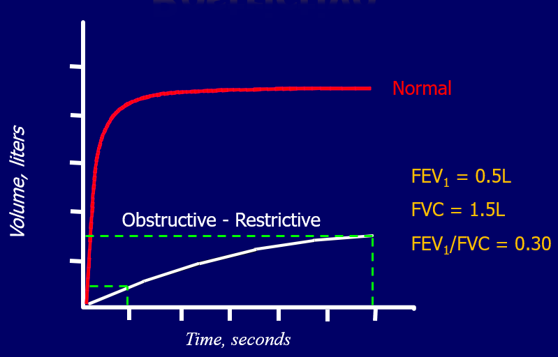 Restrictive and mixed obstructive-restrictive are difficult to diagnose by spirometry alone; full respiratory function tests are usually required
(e.g., body plethysmography, etc)