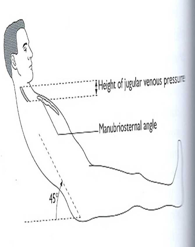
- Patient at 45 degrees
- Good lighting
- Internal jugular vein
- Reflects right atrial pressure
- Zero point = sternal angle
- Visible but not palpable
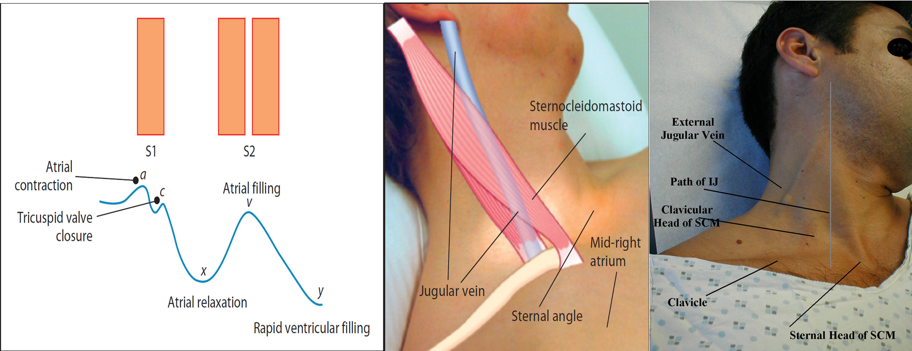
- Complex wave form (a, c, v waves)
- Decreases on inspiration
- Fills from above
- Hepatojugular reflux
- Abnormal if >3 cm above zero point:
- RV failure
- RV infarct
- Tricuspid stenosis
- Tricuspid regurgitation
- Pericardial effusion
- SVC obstruction
- Fluid overload
Note
Observe the height of JVP when patient is in the bed at 45o
Access vertical height in centimeters above the sternal
angle (normal 2-4cm)
Observe the character of JVP
- Look for a-wave (Atrial contraction)
- v-wave (Atrial filling when tricuspid wall is closed)
Large a-waves are caused by - Tricuspid stenosis - Pulmonary stenosis - Pulmonary hypertension
Important - Absent a-wave in Atrial fibrillation
Large v-wave - Tricuspid incompetence


Jagular venous pressure (JVP).
- Position of the patient at (45), head.
- Features of JVP, how to differentiate from carotid pulsation in the neck.
- Hepato-jugular reflux’ .
- Waves.
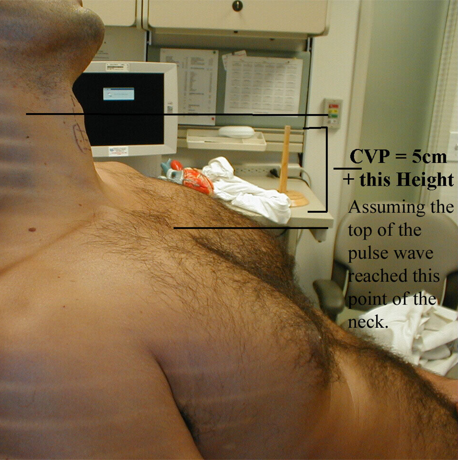
JVP = 5cm (height sternal manubrium jxn is above RA) + vertical distance from sternal manubrium jxn to top of pulse wave
•Normal < 8 cm
