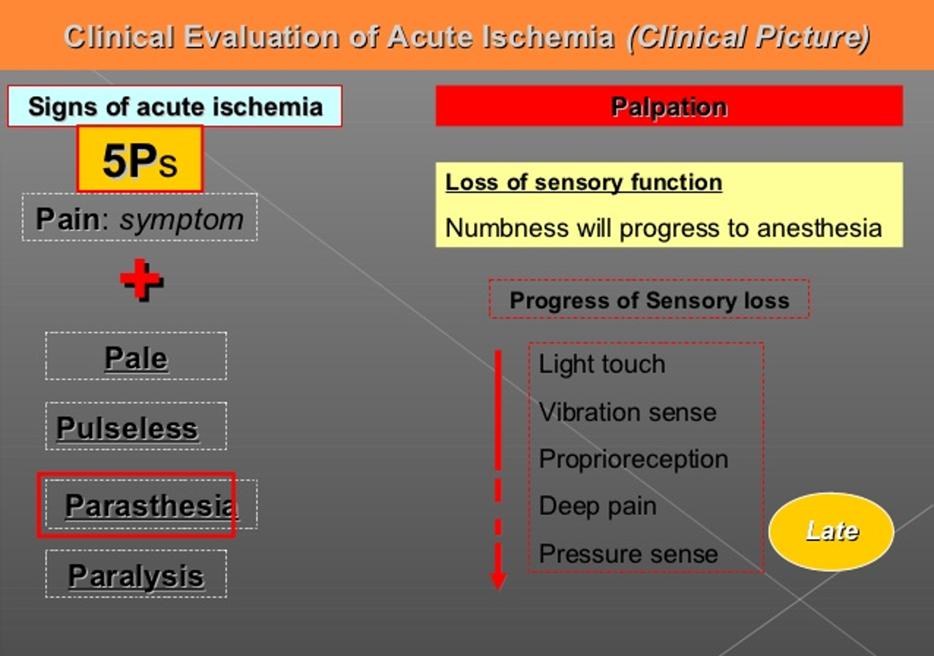
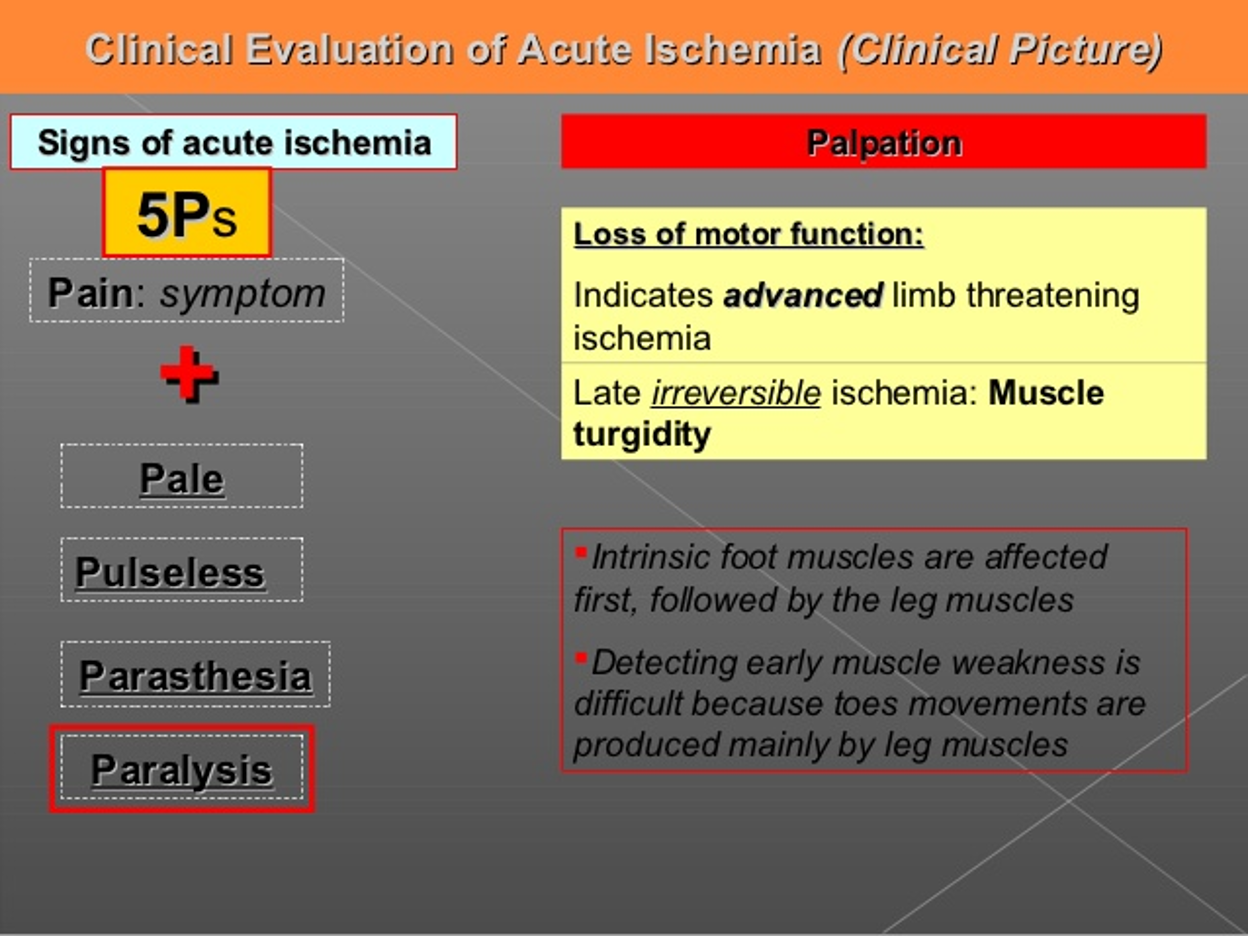
 Early -⇒ Late
light touch ⇒ vibration ⇒ Proprioreception ⇒ Deep pain ⇒ Pressure sense
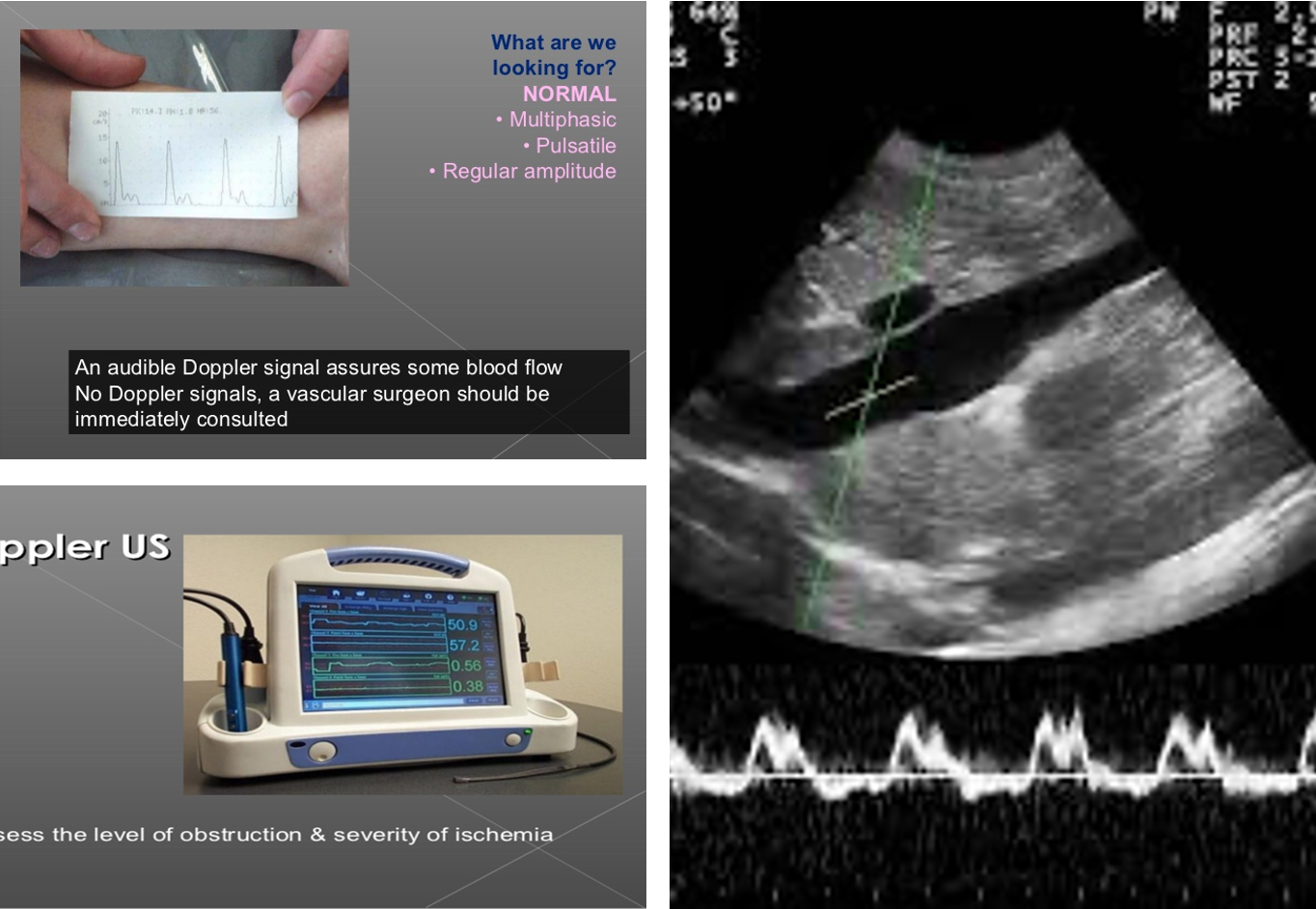
Doppler Vs Dopplux
Multiphasic normal one -
duplex waves + picture
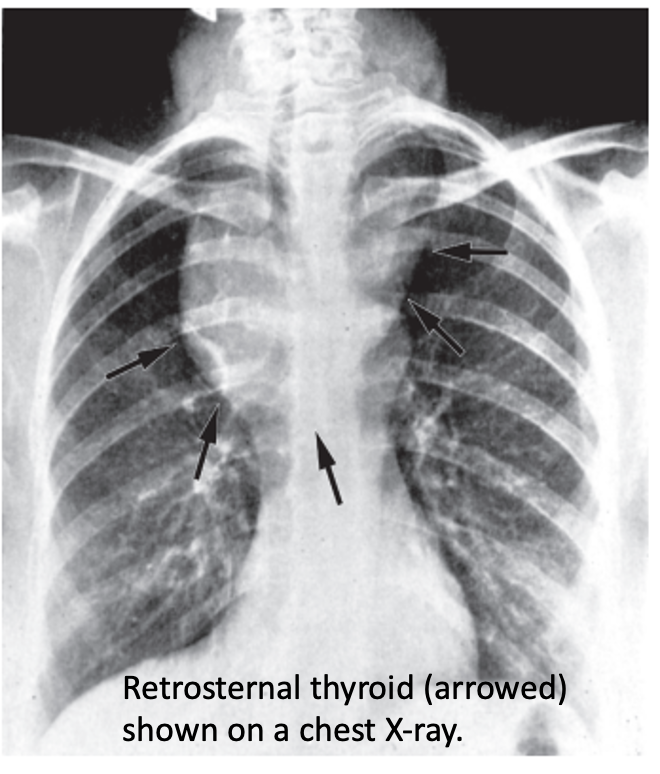
 Pemberton’s sign is used to evaluate venous obstruction in patients with goiters. The sign is positive when bilateral arm elevation causes facial plethora. It has been attributed to a “cork effect” resulting from the thyroid obstructing the thoracic inlet, thereby increasing pressure on the venous system.
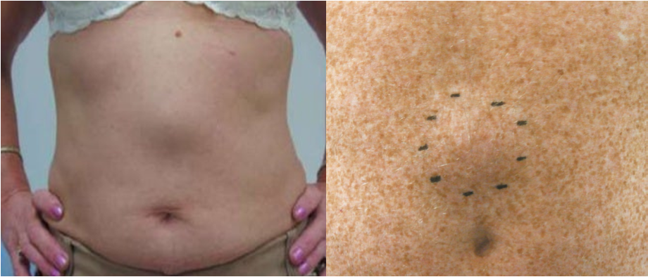
Epigastric hernia
- Protrusion through a defect in linea alba, Firm midline lump.
- Often contains preperitoneal fat or peritoneal sac with omentum
- Open surgical repair by non-absorbable suture or mesh
- Laparoscopic repair- if large
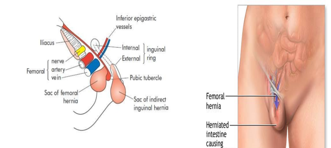
Femoral hernia
- Projects through femoral ring and passes down the femoral canal (1.25 cm)
- Appears, to lie in front of inguinal ligament
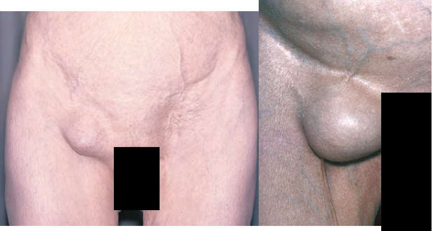
Clinical features of Femoral hernia
- Groin swelling - often small
- Groin pain on exercise
- Often irreducible due to its curved course
- Sometimes difficult to distinguish with IH
- Examination: Put a finger tip over pubic tubercle (How to find it?) IH- above & medial FH- below & lateral
- Obstruction, strangulation rate high (40%)

Differential diagnosis- Femoral hernia
- Lymphadenopathy : Cloquet’s node
- Saphenous varix- thrill on cough, disappears on lying down
- Inguinal hernia
- Ectopic testis
- Psoas abscess
- Femoral aneurysm
- Soft tissue neoplasms as lipoma
more desposed to strangluation
Incisional hernia
- Hernia through poorly healed abdominal incisions
- More common with midline vertical incisions
Predisposing factors:
- Poor surgical technique: Layered closure, absorbable suture, suturing under tension, drain tubes are brought out through the main wound
- Preoperative straining factors: Chronic cough, chronic constipation and urinary obstruction.
- Postoperative complications: Abdominal distension, cough, respiratory distress due to pneumonia or lung collapse, and postoperative wound infection.
- General factors: Age (elderly patients), malnutrition, hypoproteinemia, jaundice, malignancy, diabetes, chronic renal failure, steroid or immunosuppressive therapy and alcoholism.
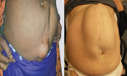
Gastric Adenocarcenoma:
- 95% of all malignant gastric neoplasms
- Twice as common in men as it is in women,.
- Increases with age, peaking in the seventh decade.
- More at gastric cardia
Risk factors:
- Nutritional
- Environmental
- Social
- Medical

- left supraclavicular lymph virchow sign
- , 3, 4 sister mary joseph nodules
S/S
-
Significant GI bleeding is rare
-
15% of patients may develop hematemesis,
-
40% of patients are anemic.
-
Physical signs develop late & associated with locally advanced or metastatic disease.
-
Palpable abdominal mass,
-
Palpable supraclavicular (Virchow’s)
-
Periumbilical (Sister Mary Joseph’s) lymph node,
-
Peritoneal metastasis palpable by PR (Blummer’s shelf),
-
Palpable ovarian mass (Krukenberg’s tumor).
Plain x-ray- CXR, AXR, tomograms
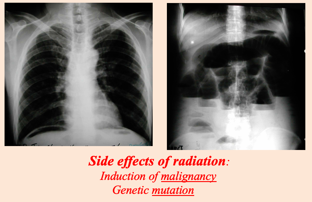 Modality: plain x-ray
Findings: air under diaphram / multiple air fluid levels
Differentials: perforation / obstruction
Side effects of radiation:
- Induction of malignancy
- Genetic mutation
Subcutanous emphysema
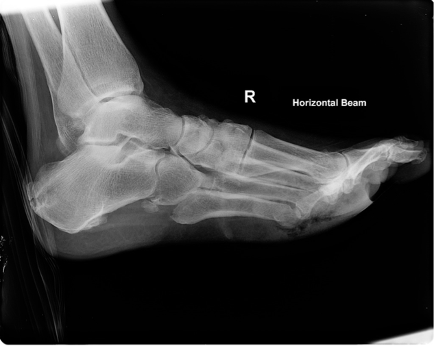
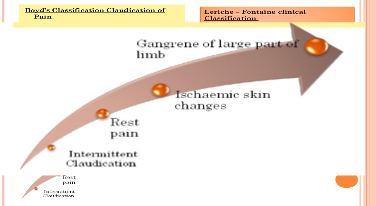 Intermittent claudications ⇒ Rest pain ⇒ Ischemic skin changes ⇒ gangrene Large part of limb
Deep Vein Thrombosis:
-
- Post-operative.
-
- Immobility due to other illness.
-
- Leg pain.
-
- Leg swelling.
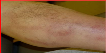
Superficial thrombophlebitis:
- Inflammation & thrombosis of previously normal superficial vein.
- Pain, redness and cord like vein
antibiotic enough
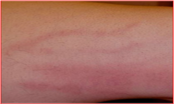
Venous ulcer:
- Previous DVT , Varicose Veins
- Above medial malleolus (70%)
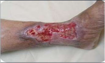
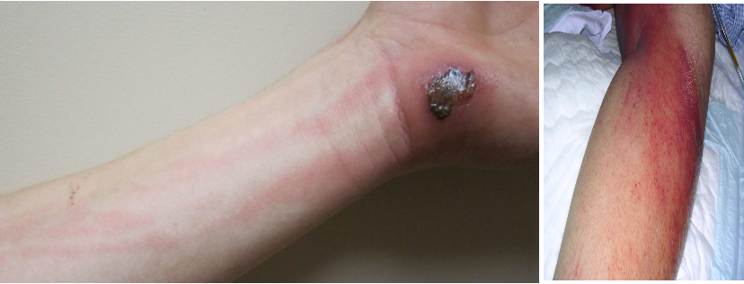
Infection- lymphangitis
Inspection:
- Red streaks and swelling of the limb
- Site of primary infection may be visible
- Spreading
Palpation:
- Warm, tender, pitting oedema
- Palpable and tender draining lymph node
Lymphedema
- Primary lymphedema: Congenital, due to poorly developed lymphatics
- Secondary: Infective (Filariasis) or neoplastic (secondary deposits)
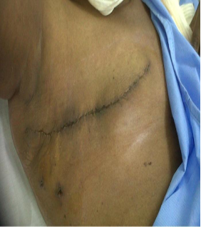
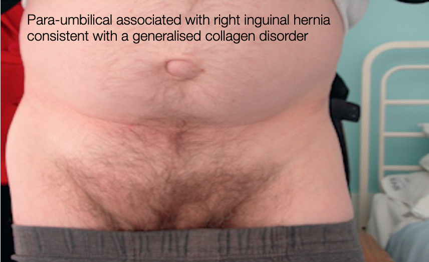
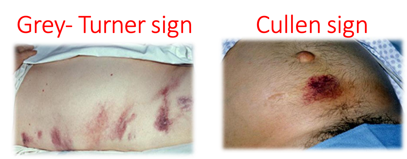
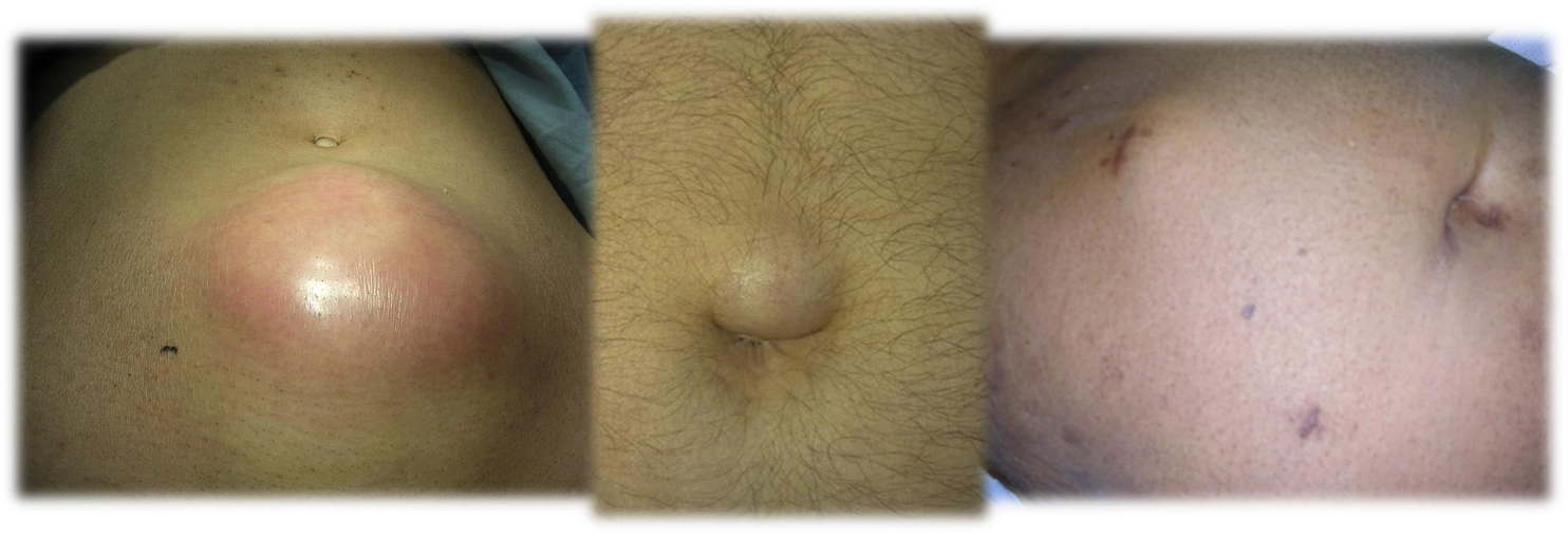 1- abcess 2- periumbilical hernia 3- inisional hernia
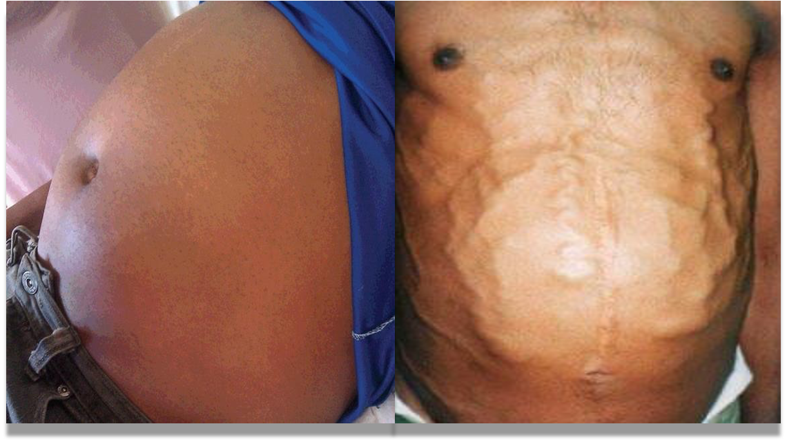 ascites / caput medusa
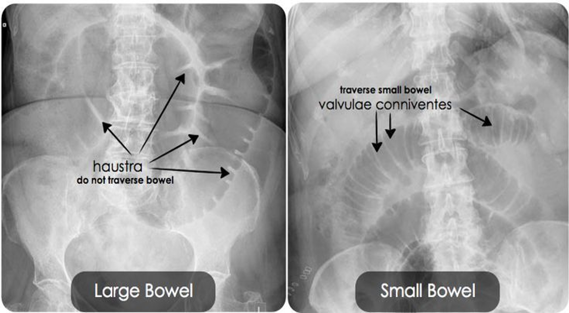
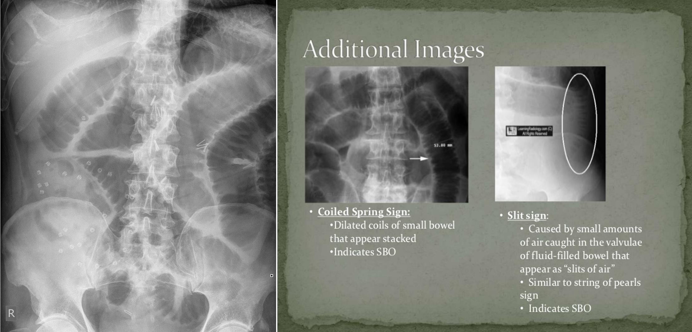
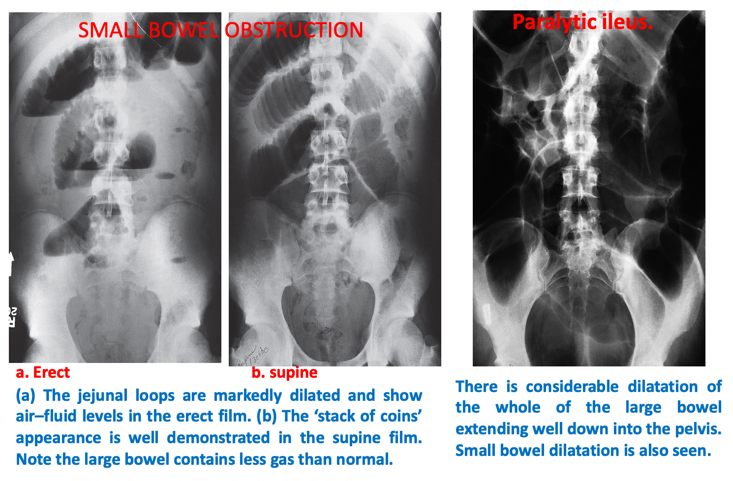
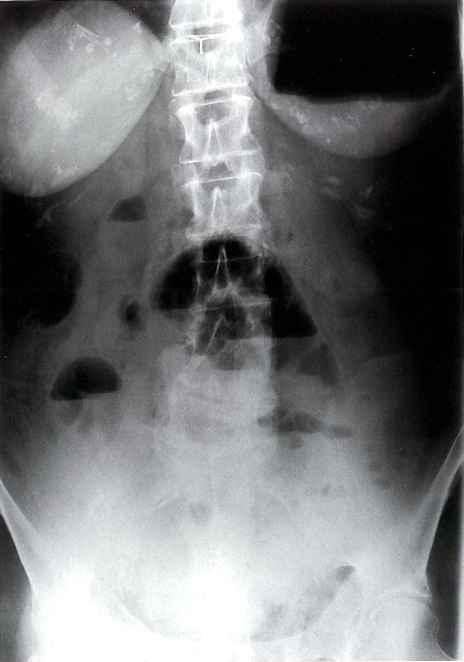 Mechanical small bowel obstruction-X-ray abdomen (erect)
Multiple air-fluid levels are visible in the mid-abdomen. The opaque appearance of the pelvis is due to fluid-filled loops of small bowel. There is a paucity of gas in the colon, and an air-fluid level is present in the dilated stomach.
