Internal Medicine
DR WAQAR
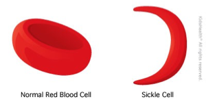
NORMAL
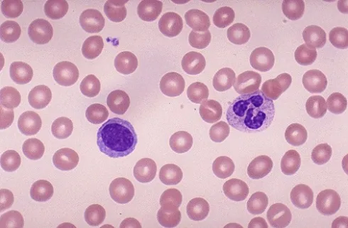
- RBCs are round
- Most of the hemoglobin is Hb.A
WHAT IS SICKLE CELL DISEASE
- It is a type of hemoglobinopathy
- It is a hereditary condition
- An abnormal Hb. called Hb.S, replaces the normal Hb.A
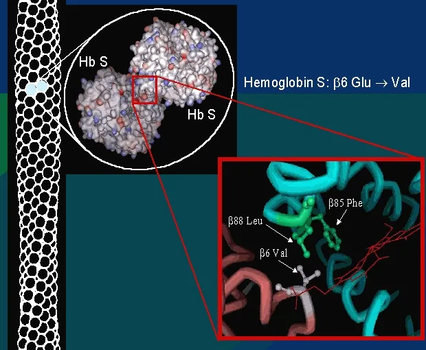
- Hb.S is produced when the amino acid “glutamic acid” (in the normal Hb.A) is replaced by another amino acid “valine”
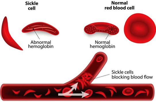
- The Hb.S causes the RBCs to become sickle-shaped (MANJAL) which causes problems (blocking of blood vessels & hemolysis)
VASO-OCCLUSION BY SICKLED CELLS
GENETICS OF SCD
- Pattern of inheritance: Autosomal recessive
- Normally, Hb. is produced by 2 genes
- If both genes are abnormal → sickle cell disease (no Hb. A)
- If only one gene is abnormal, it is called sickle cell trait (carrier state, Hb.AS) → half of body’s Hb is HbS & half is Hb A. Not full-blown disease
- Normal Genetics: AA
- Sickle cell trait: AS
- Sickle cell disease: SS
VARIANTS OF SCD
- Hb.SC disease: Presence of Hb.S plus Hb.C (hemoglobin C is another type of abnormal Hb.)
- Hb.S/β: Sickle cell gene plus beta-thalassemia gene
- Sickle cell disease (Hb.SS) → Symptomatic
- Sickle cell trait (Hb.AS) → usually asymptomatic but can cause sudden death if patients do vigorous exercise
- Hb.S/β disease (sickle + thalassemia) → Symptomatic
- Hb.SC disease: Symptomatic, but milder than SCD
EPIDEMIOLOGY
- Very common worldwide
- In the U.S., 1 in 625 births have SCD
- More in African-Americans
- Common in Mideast, Africa, Mediterranean areas (Greece, Turkey, Italy)
- In KSA, high prevalence in Eastern region & southwestern areas (Jizan)
SIGNS & SYMPTOMS
A stable patient (no acute symptoms) will have:
- Anemia (Hb. around 8)
- May be jaundice (due to?)
- Sometimes leg ulcers
Various Crises in SCD
- Vaso-occlusive pain crisis
- Acute chest syndrome
- Aplastic crisis
- Pain Crisis
- Hemolytic crisis
- Splenic sequestration crisis
OTHER FEATURES OF SCD
- Priapism: It is a painful erection of male genitalia. Repeated attacks may cause impotence. Treatment: analgesia, transfusion
- Stroke/TIA: Due to blockage of cerebral vessels.
- Asplenia (non-functioning spleen)
- Repeated vaso-occlusion causes splenic infarcts → spleen shrinks & becomes non-functional
- Most cases occur by the age of 1 year
- Non-functioning spleen → risk of infections with encapsulated bacteria (Pneumococci, meningococci, H.Flu.)
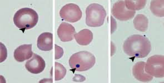
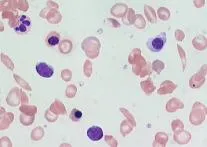
- Howell-Jolly bodies seen on a peripheral smear (remains of DNA in the RBCs) (Howell Jolly bodies indicate asplenia)
(Howell-Jolly bodies seen if there is no spleen)
-
Renal problems:
- Renal medullary infarction → causes tubular damage → impaired water absorption → fluid loss
-
Retinopathy: Due to retinal infarcts (vaso-occlusion)
-
Cholelithiasis: Hemolysis → increased bilirubin → bilirubin stones in the gallbladder (look for a surgical scar)
-
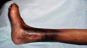
Leg ulcers:
- Due to vaso-occlusion
- Usually on the ankles
-
Pulmonary HTN: Increased risk of this in SCD patients.
-
Bone problems:
- Increased fractures
- Osteomyelitis: Due to Staph Aureus (most common) & Salmonella (most severe)
- Avascular necrosis of femoral & humeral head (due to vaso-occlusion)
-
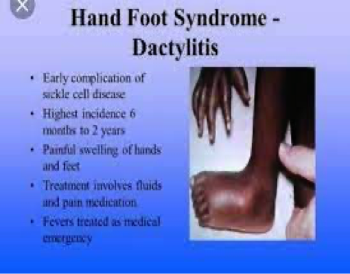
Dactylitis:
- Pain & swelling in the fingers & toes
- Often the presenting feature in children
- Due to ischemic damage to the bones
- Also called HAND-FOOT disease
SCD Investigations
CBC, LFT, Peripheral smear, Electrophoresis, Solubility test, sickling test, genetic testing
Treatments for SCD
Longterm
- Hydroxyurea,
- Folic acid tablets,
- L-Glutamine (oral)
Other Treatments
- Stem cell transplant
- Gene therapy
Rapid Fire Questions
- What is the inheritance pattern of SCD? autosomal recessive
- What is the genotype of a normal person, SCD, and trait? SS fullblown, S trait
- Which amino acid is present in SCD? valine
- Is sickle cell trait symptomatic? usually asymptomatic
- CBC findings in SCD? high plates
- Why is retic count high? hyperactive marrow
- Name some specific tests to diagnose SCD? electrophoresis
- What is the cause of jaundice in SCD? hemolysis
- Name 5 acute crises in SCD? aplastic, splenic, …
- Transfusion is the main treatment of pain crisis, right or wrong? wrong
- What is the cause of pain in acute pain crisis? vasoblockages; ischemic
- 5 indications of transfusion in SCD? HASCS
- What can precipitate a pain crisis? infection, stress
- Treatment of pain crisis? transfusion analgesics
- What is the cause of aplastic crisis? bone marrow suppression due parrovirus b
- What will CBC show in aplastic crisis? everything is low
- Treatment of acute chest syndrome? transfusion, antibiotics, analgesics
- Name 3 crises which can cause a sudden Hb drop? aplastic, hemolytic, splenic…
- What happens to the spleen in SCD patients? fibrosis; aspleenia
- What happens to the eyes, male genitalia, brain, kidneys, bones? retinopathy//occlusions .. .. >
- Patient with SCD has a surgical scar in the right upper abdomen. Why? cholethiasis
- What vaccines should be given to SCD patients? infections that perticipates due asplenia
- In children with SCD, what can happen in the hands & the feet? dactytilitis
- Name 3 medicines which are given long term? folic acid, L-glutamine, Hydroxyurea
- What preventive measures should be taken in SCD patients? prophylaxis, dont go abha, take antibiotics when fever + respiratory distress
- In a patient with SCD, what will you see in a peripheral smear? sickle
- High chances of infection with which bacteria? encapsulated; asplenia
- Name 2 advanced treatments for SCD? stemcells & Gene Therapy
Pediatrics
“Sickle cell disease (SCD) is a term used for a group of genetic disorders characterized by production of Hb “S”. Sickle cell hemoglobinopathy occurs due to mutation of beta-globin gene situated on short arm of chromosome 11, where adenine is replaced by thymine in base of DNA coding for the amino acid in the sixth position in beta-globin chain.
Pathophysiology
-
The “sol” (soluble) form of Hb changes to “gel” form, when Hb “S” is deoxygenated. ⇒
-
In gel form the Hb crystallizes to small, rigid, boat-shaped objects known as ‘’tactoids’’. These tactoids polymerize forming insoluble structure, deforming the RBC to sickle shape. ⇒
-
The RBC membrane becomes more fragile. ⇒
-
The polymerization is facilitated by increased concentration of Hb “S”, acidic pH, low oxygen saturation, stasis of blood flow, decreased levels of Hb”F”.
-
Upon reoxygenation - The sickle cell initially resumes normal configuration, but with repeated cycles of sickling and unsickling, fixation of membrane occurs in sickled configuration, leading to irreversible sickle cell formation, and hemolysis. ⇒
-
Platelets aggregate over the adherent red cells and damaged endothelium, causing blockage of microvasculature and ischemia of the tissue.
-
Infarction of liver, spleen, muscles & bones. (Hepatomegaly & Jaundice, abd. Pain & fibrosis & ↓ spleen size, bone & joint pain).
- Sickle cells sequestered in RE cells and are hemolysed → Anemia
- Chronic Hypoxia → - Clubbing of fingers & toes - Chronic Leg ulcer - Growth retardation
- ↑Bilirubin turnover → Pigment stones
Various Crises in SCD
- Vaso-occlusive pain crisis
- Acute chest syndrome
- Aplastic crisis
- Megaloblastic Crisis
- Pain Crisis
- Hemolytic crisis
- Splenic sequestration crisis
Stroke
- Common age group is 3–10 years. Around 70–90% patients experience repeated episodes of stroke within 36 months unless they are on transfusion regimen maintaining Hb”S” level below 30%.
Priapism
- Priapism is persistent painful penile erection. Recurrent acute episodic attacks last from few minutes to several hours. It usually subsides spontaneously. Impotency may be a sequel. Blood transfusion is indicated if pain, engorgement persists for 24–48 hours.
Clinical Features
- Moderate to severe anemia.
- Mild jaundice.
- Non-healing leg ulcers.
- Decreased physical growth & delayed puberty.
- Clubbing of fingers & toes.
- Enlarged heart and hemic murmur.
- Hepatomegaly.
- Splenomegaly in the beginning.
- Sickle cell crisis –
- Pains & aches.
- ↑ jaundice.
- ↑ anemia.
- Fever.
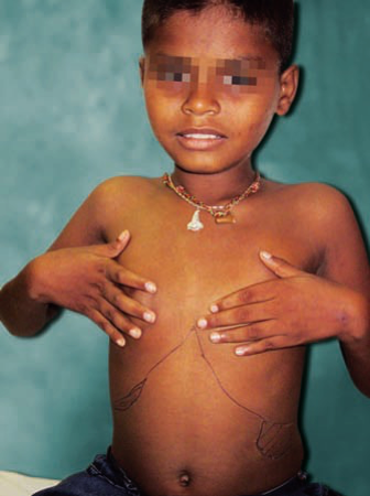
Complications: Z
- Bones → Dactylytis and chronic osteomyelitis, ⇒ Aseptic necrosis of head of femur.
- Eyes → Sudden blindness.
- Brain → Neurological deficits & emotional disturbances.
Diagnosis Z
- Screening test of older children can be done sickling test.
- Diagnosis can be confirmed by Hb electrophoresis, globin DNA analysis.
- Antenatal diagnosis can be done at 8–10 weeks of gestation, with chorionic villus biopsy, or amniocentesis. DNA analysis using PCR.
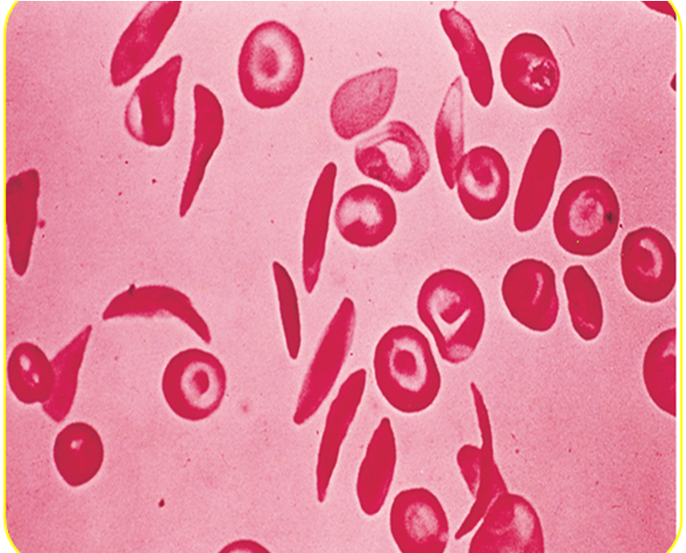
Peripheral smear:
- Deformed sickle-shaped red cells.
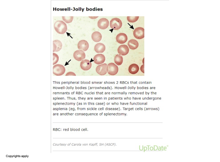 The image shows a microscopic view of a peripheral blood smear. In this image, there are red blood cells (RBCs) with specific annotations pointing out Howell-Jolly bodies (indicated by arrowheads) and target cells (indicated by arrows). Howell-Jolly bodies are remnants of RBC nuclei usually removed by the spleen. They are observed in patients who have undergone a splenectomy or have functional asplenia, such as from sickle cell disease. Target cells can also occur following a splenectomy.
Treatment
Painful Crisis
- Mild pain: codeine, aspirin, ibuprofen, acetaminophen, naproxen.
- Severe pain: meperidine, tolmetin, oxycodone, Ketamine.
- Vasodilators: Pentoxifylline, nifedipine, and buflomedil increase microvascular circulation.
- Hydration.
- Blood transfusion: may be appropriate if pain is accompanied by another complication such as stroke or ACS.
- Hydroxyurea: leads to increase in Hb “F”, which ameliorates the severity of SCD.
Newer Treatments
-
Agents stimulating Hb F - Hydroxy urea - 5 – aza cytidene - Butyrates
-
Agents ↓ Hbs concentration - DDAVP: a synthetic version of antidiuretic hormone (used in von willebrand’s disease to stimulate vWF, may also be used in mild hemophilia A) - Nifedepine, verapamil
-
Membrane active agents – Zinc
-
Hb solubility increasing agents - Cyanate - Urea - Phenyl alanine
A life-long cure for SCD is available only through hematopoietic stem cell transplantation.
Prevention Z
- Avoid precipitating factors for crisis - Exposure to hypoxia, dehydration, extreme cold, extreme heat, change in altitude, infections.
- Avoid Dehydration and loss of electrolytes may lead to red cell dehydration precipitating sickling process.
- Parent’s education.
- Immunization: pneumococcal and Influenzae (Hib) vaccine. Hepatitis B vaccine.
- Hemoglobin levels should be maintained at 10–12 g/dl.
- Folate supplementation.
- Genetic counseling and mass education of high-risk.
- Oral penicillin prophylaxis. Oral penicillin should be given to children up to the age of 5 years.
FM
Jazan Najran Z
Sickle Cell Anemia
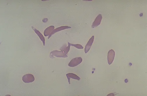
Sickle Cell Anemia
- An inherited disease of red blood cells
- Affects hemoglobin
- Polymerization of hemoglobin leads to a cascade of effects decreasing blood flow
- Tissue hypoxia causes acute and chronic damage
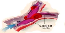
Why Do Cells Sickle?
- Glutamic acid is substituted for valine
- Allowing the polymerization of sickle hemoglobin when deoxygenated
Normal Vs. Sickle Red Cells
Normal
- Disc-Shaped
- Deformable
- Life span of 120 days
Sickle
- Sickle-Shaped
- Rigid
- Lives for 20 days or less
Hematology Overview
Pathology
Hemolysis
-
The anemia in SCD is caused by red cell destruction, or hemolysis, and the degree of anemia varies widely between patients.
-
The production of red cells by the bone marrow increases dramatically, but is unable to keep pace with the destruction.
Vaso-occlusion
-
Occurs when the rigid sickle-shaped cells fail to move through the small blood vessels, blocking local blood flow to a microscopic region of tissue.
-
Amplified many times, these episodes produce tissue hypoxia.
-
The result is pain, and often damage to organs.
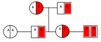
Sickle Cell Pedigree
- Parents with sickle cell trait: hemoglobin AS
- Probability of child with hemoglobin AA: 25%
- Probability of child with sickle cell trait AS: 50%
- Probability of child with sickle cell disease SS: 25%
Clinical Manifestations
- Thrombotic crises
- Aplastic crises
- Hemolytic crises
- Sequestration crises
Laboratory Findings
- Anaemia and unconjugated hyperbilirubinemia
- Sickling test: positive
- Hb electrophoresis: Hbs and HbA2 and HbF but not HbA
Treatment
- Blood transfusion in severe cases and aplastic crises
- Infection prevention
- Pain management
- Maintenance of hydration
- Chronic disease management
- Splenectomy if the spleen is huge

Radiology
Acute clinical manifestations
- Vaso-occlusive events
- Dactylitis in children < 5 years of age
- Vasoocclusive crises (sickle cell pain crisis) Acute chest syndrome
- Organ infarctions (any organ; particularly the spleen) It is well demonstrated on CT
- Avascular necrosis
Infection
- Pneumonia
- Osteomyelitis (most common cause: Salmonella;Staph Aureus)
- Acute hemolytic crisis
MSK imaging features of SCD:
-
Bone infarction: Occur in the intra-articular portions of the bones (avascular necrosis ) or in the medulla of a bone
-
Hair-on-end sign
-
Evidence of bone infection.
-
H-shaped vertebrae
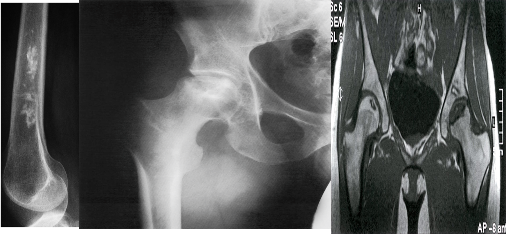 A) Bone infarct. There is calcification in the medulla of the lower end of the femur.
B) X-ray right hip (AP view) The articular surface of the femoral head is irregular (black dotted line) and the underlying bone is heterogeneous (green overlay) as a result of osteonecrosis. A zone of sclerosis (red overlay) is present along the interface with normal bone.
C) Bilateral osteonecrosis of the femoral head
MRI hips (T1 weighted; coronal view) of a 32-year-old woman with groin pain
There is a well-defined crescent-shaped area of intermediate to low signal within the subchondral marrow of the left femoral head . A thick peripheral band of even lower signal separates this region from the hyperintense normal bone marrow. On the right side, there is a slightly smaller area of abnormal oval-shaped signal within the epiphysis of the right femoral head.