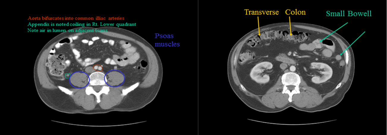
CT can show the full width of the wall of the structures in question as well as the surrounding fat.
In addition, the lumen of the gastrointestinal tract may be evaluated using Gastrografin as the contrast agent.

Hepatobiliary Computed tomography
Hi The normal hepatic parenchyma has a relatively high density prior to contrast enhancement; higher than that of muscle and higher or equal in density to the spleen.
On images taken without intravenous contrast medium, the hepatic veins and portal veins are seen as branching, low density structures coursing through the liver.
The normal intrahepatic bile ducts are not visible.
Intravenous contrast medium is usually given in order to increase the density of normal liver parenchyma and to emphasize the density difference between the normal parenchyma and lesions that enhance poorly, such as tumours or abscesses.

-
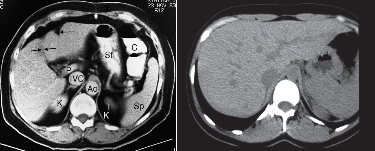
CT scan of normal liver (enhanced scan). Ao, aorta; C, colon; IVC, inferior vena cava; K,kidney; P, portal vein; Sp, spleen; St, stomach. The single arrow indicates a fissure for the falciform ligament, and the double arrows a fissure for the gall bladder, which divides the liver into the right and left lobes.
-
in second image CT scan of normal liver showing unopacified veins, which should not be confused with metastases.
Phases of liver opacification by contrast
Scanning during the arterial phase: About 30 seconds after the injection of contrast, will show lesions such as haemangiomas and some neoplasms, particularly hepatomas and highly vascular metastases (e.g. carcinoid), as areas of greater enhancement than the surrounding parenchyma.
Portal venous phase : Scan taken 60–70 seconds after the injection of contrast. Most metastases are best demonstrated as low attenuation areas during portal venous phase. It is the best for evaluating the solid organs in the abdomen
Delayed phase : Scan taken 6 to 10 minutes after contrast injection