Infantile hemangioma (IH)
Infantile hemangiomas (IH) are common benign vascular tumors of infancy occurring in about 4% of infants
At birth, a premonitory mark may be present such as a bruise-like patch, area of vasoconstriction/pallor or telangiectasias
Risk factors for developing IH
- Low birth weight
- Female gender
- Twin gestation
- Fair skin
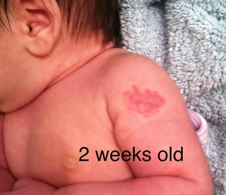
IH at 2 weeks old
-
Infantile hemangiomas proliferate during the first 2-3 months of life
-
Growth usually stabilizes around 4-6 months, followed by involution over years
-
Larger, deeper IH grow for longer and involutes more slowly.
-
Based on these growth characteristics, IH requiring intervention should be referred for treatment before 3 months of age
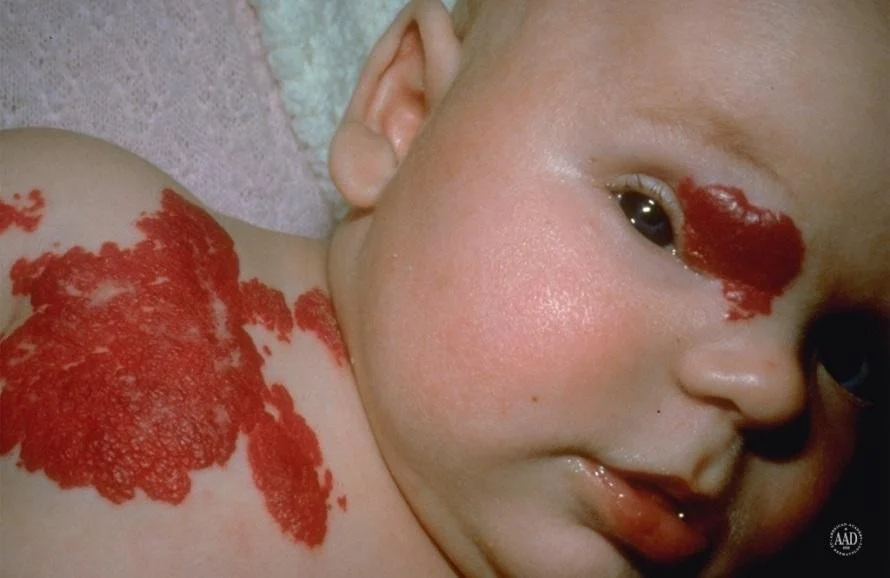
Types of heamangiomas
- Superficial
bright red and minimally elevated

- Deep
large with a bluish color

- Mixed
have both components

Hemangiomas: clinical appearance
Hemangiomas may occur anywhere on the body
They may be localized or segmental/regional in distribution
![[Infantile hemangioma IH-1747879722537.webp]]
Complications include:
- Ulceration
- Visual Impairment
- Airway involvement
- Multifocal presentation
- Aesthetic complications
- Complex Associations
Infantile Hemangiomas: Management
Systemic Therapies
- Oral propranolol
- Systemic corticosteroids
- Immunosuppressive or anti-neoplastic** therapies (Interferon, Vincristine)
Local Therapies
- Topical corticosteroid s
- Topical beta blockers
- Intralesional corticosteroid s
- Pulsed dye laser for ulceration or residual lesions
Clinical Case
HPI: This is an 8 day old baby boy who presents for evaluation of a large, pink mark on his forehead present at birth.
His parents are concerned by the size and location of the birthmark.
PMH: Born full term to healthy mother, no pregnancy or labor complications, received all vaccinations
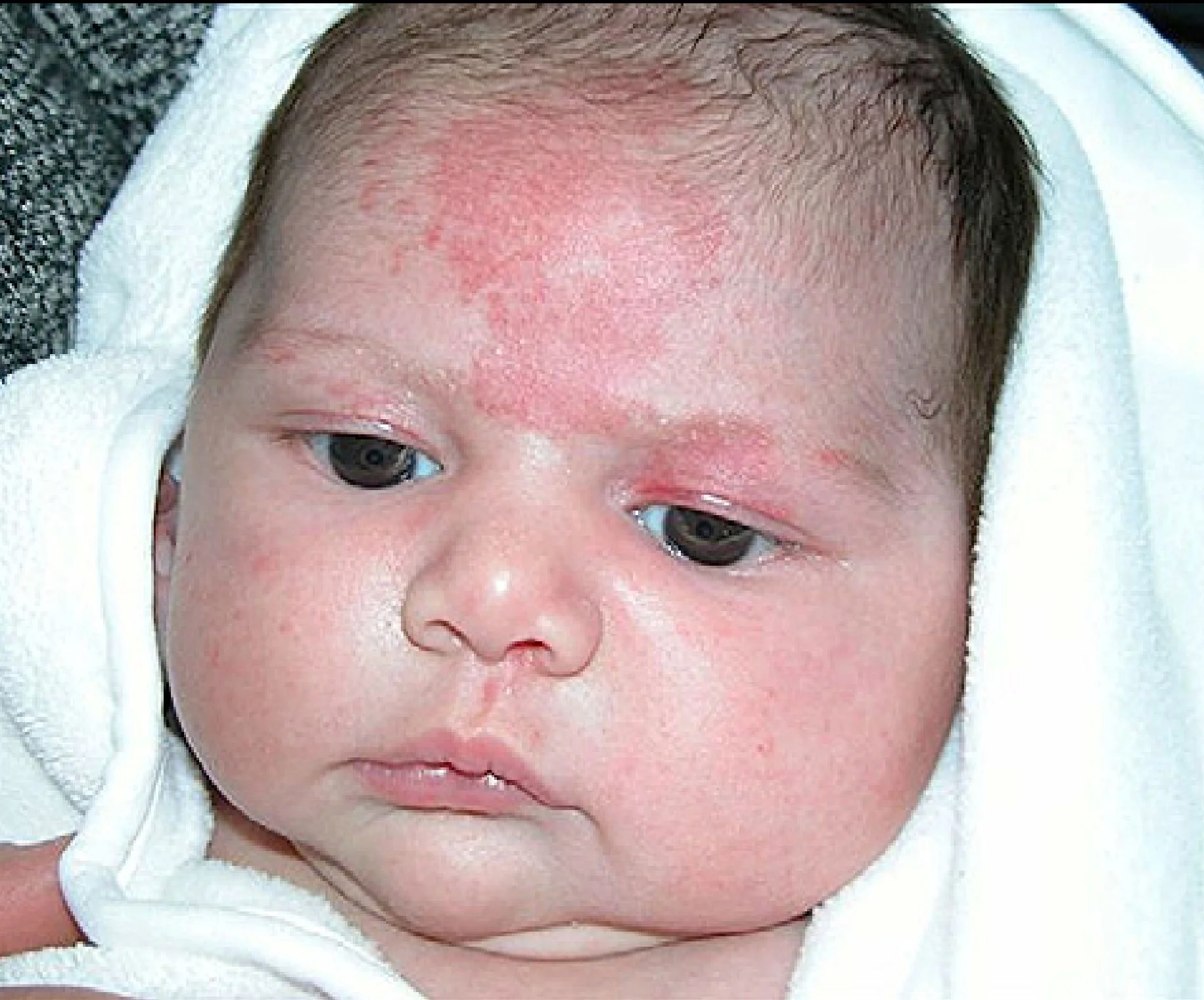 Irregular well defined erythematous patch in forehead