Omar Alaidaroos
Diseases of small bowel
-
Bowel obstruction
- Mechanical vs. adynamic
- Acute vs. chronic
- Open V s Closed
- Partial vs. complete
-
Paralytic Ileus
-
Mesenteric Ischemia
-
Crohn’s Disease
-
Meckel’s diverticulum
-
Intussusception
-
Small bowel neoplasm
-
Small bowel lymphoma
-
Short gut syndrome
Anatomy
- Small bowel extends from the pylorus to the ileocaecal junction.
- 2.75 -10.49 m long, & 2.5–3 cm in diameter.
- The surface area of its mucosa is 30 square meter
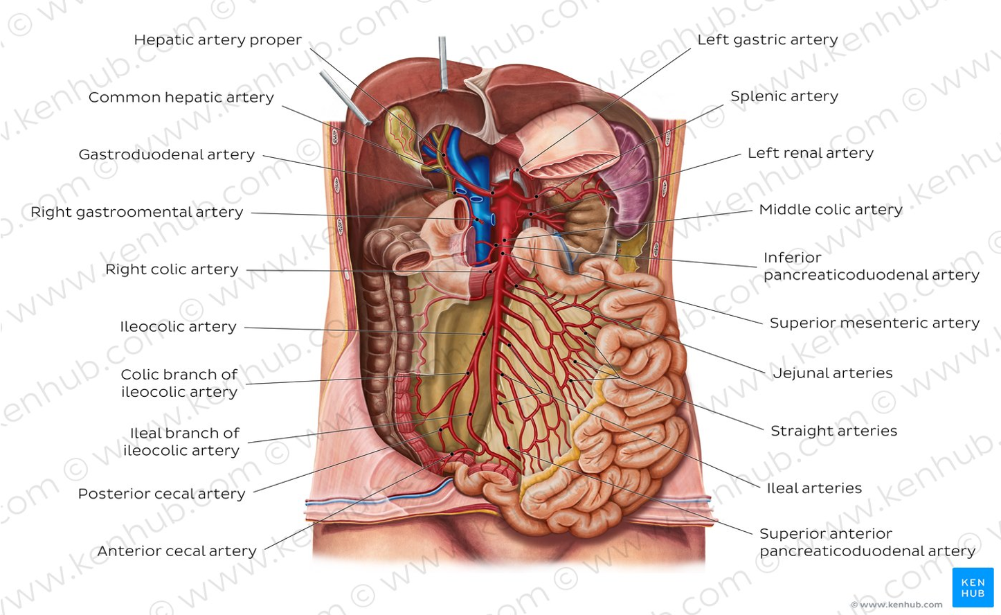
- It receives blood supply from the coeliac trunk, & superior mesenteric artery
- 3 distinct regions – the duodenum, jejunum, & ileum.
- Ileocecal sphincter – Transition between small and large intestine
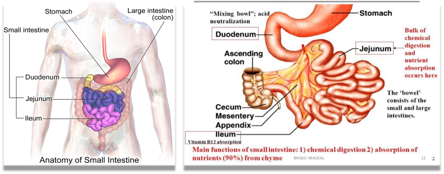
Function:
- Absorption of nutrients, & minerals from food, using small finger-like protrusions called villi.
- (sugars, amino acids, and fatty acids)
- Chemical digestion
- Absorbed in Duodenum → Fe, Ca++, B2, B6.
- Absorbed in Jejunum → B1 & B9.
- Absorbed in terminal ileum → B12, bile salts, Vits ADEK
Parts of small intestine
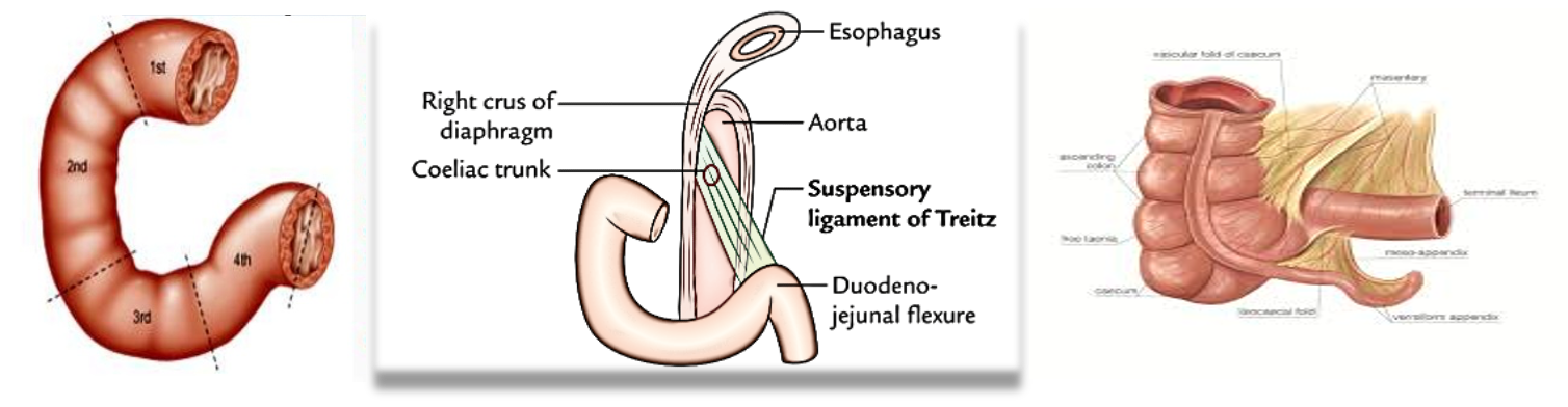
Duodenum:
- Shortest part (20-25 cm) in length, & preparation for absorption begins.
- Shaped like a “C”. It surrounds the head of the pancreas.
- It has 4 parts
- Receives bile through CBD, and pancreatic juice through
- the pancreatic duct, controlled by the sphincter of Odd
- It has Bruner’s glands, which empty into the intestinal glands, secrete an alkaline fluid composed of mucin, which exerts a physiologic anti-acid function by coating the duodenal epithelium, therefore protecting it from the acid chyme of the stomach.
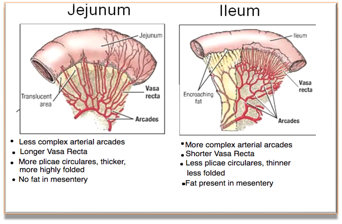
Jejunum:
- Is the midsection of the small bowel, connecting the duodenum to the ileum.
- It is about 2.5 m long
- Contains the plicae circulares, & villi that increase its surface area.
- The suspensory muscle of the duodenum marks the division between the duodenum and the jejunum.
Ileum:
- It is about 3 m long and contains villi like the jejunum.
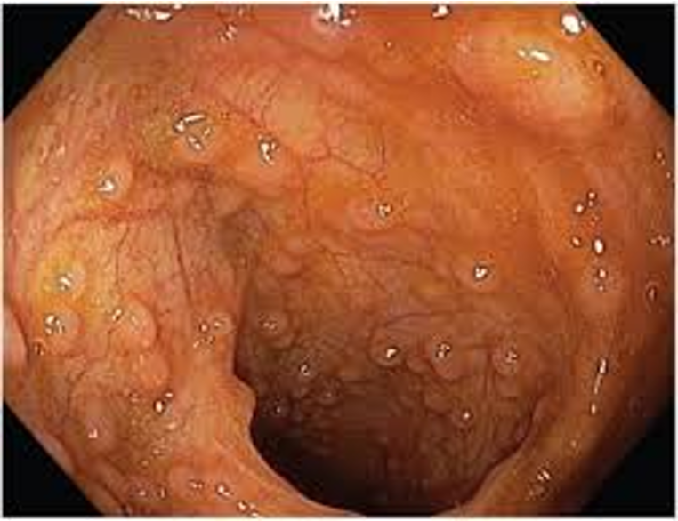
- It has Peyer’s patches (Aggregated lymphoid nodules)
- It joins to the cecum of the large bowel at the ileocecal junction
Peyer’s Patch